BIOLOGY Volume 3 - A Guide to General Biology - 2004
24. VARIATION AND GENETICS
24.6. Sex Determination
A particularly striking example of the aforementioned relationship between the phenotypic traits of organisms and their Chromosome Structure is Sex Determination. In Drosophila, phenotypic differences between the two sexes are clearly linked to differences in Chromosomes (Fig. 24.16). When studying the karyotype of males and females in A number of animals, certain differences were discovered. Both male and female individuals have pairs of identical (homologous) chromosomes in all Cells, except for one pair where they differ. These are the sex chromosomes (heterosomes). All other chromosomes are called autosomes. As can be seen in Fig. 24.16, Drosophila has four pairs of chromosomes. Three pairs (II, III, and IV) are identical in both sexes, but pair I, which consists of identical chromosomes in the female, differs from pair I in the male. The chromosomes of this pair are called the X and Y chromosomes; the female genotype is XX, and the male genotype is XY. Such differences between sex chromosomes are characteristic of most animals and humans (Fig. 24.17). In birds and butterflies, the reverse is observed: females have XY chromosomes, and males have XX. In some insects, such as orthopterans, the Y chromosome is entirely absent, so that Male Germ Cells carry a single X chromosome (male genotype XO).
Class="center">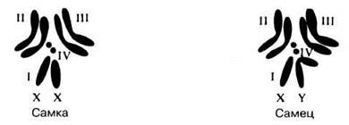
Fig. 24.16. Chromosomes of male and female Drosophila melanogaster. Four pairs of chromosomes are shown. Pair I represents the sex chromosomes.
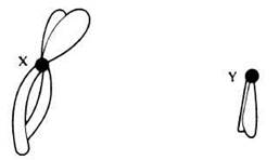
Fig. 24.17. Human sex chromosomes in metaphase of Meiosis.
During gametogenesis, typical Mendelian segregation of sex chromosomes is observed. For example, in mammals, each egg Cell contains a single X chromosome; in males, half of the sperm carry an X chromosome, while the other half carry a single Y chromosome (Fig. 24.18). The sex of the offspring depends on which sperm fertilizes the egg. An individual with the XX genotype is called homogametic, as it produces identical Gametes containing only X chromosomes, while an individual with the XY genotype is called heterogametic, since half of its gametes contain an X chromosome and the other half contain a Y chromosome. In humans, an individual's genotypic sex can be determined by microscopic examination of epithelial cells from the inner cheek. In such cells, one of the X chromosomes is in an inactive, condensed state and appears as a dense, dark body known as a Barr body. The number of Barr bodies is always one less than the number of X chromosomes present; i.e., they are completely absent in males (XY) and there is only one in females (XX). The function of the Y chromosome obviously varies depending on the animal species. In humans, the Y chromosome controls testicular differentiation, which subsequently influences The Development of reproductive Organs and the male phenotype (sec. 21.7.4). In some organisms, however, the Y chromosome contains no sex-determining genes. It is even considered genetically inert or empty because it contains very few genes. It is hypothesized that in Drosophila, genes determining male characteristics are located on autosomes, and their phenotypic effects are masked by the presence of a pair of X chromosomes. Male characteristics appear in the presence of only one X chromosome. This is an example of sex-limited inheritance (as opposed to Sex-Linked Inheritance), in which, for example, genes determining beard growth are suppressed in females.
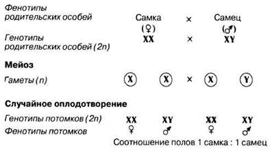
Fig. 24.18. Genetic explanation of the sex ratio in humans.
Morgan and his colleagues noticed that the inheritance of eye color in Drosophila depends on the sex of the parental individuals. Red eye color is dominant over white. When crossing a red-eyed male with a white-eyed female, the F1 generation produced an equal number of red-eyed females and white-eyed males (Fig. 24.19, A). However, when crossing a white-eyed male with a red-eyed female, the F1 generation yielded equal numbers of red-eyed males and red-eyed females (Fig. 24.19, B). Intercrossing these F1 flies produced red-eyed females, red-eyed males, and white-eyed males, but not a single white-eyed female (Fig. 24.19, C). The fact that the recessive trait appeared with a higher frequency in males than in females suggested that the recessive allele determining white eyes is located on the X chromosome, while the Y chromosome lacks the eye color Gene. To test this hypothesis, Morgan crossed the original white-eyed male with a red-eyed female from the F1 generation (Fig. 24.19, D). The offspring consisted of red-eyed and white-eyed males and females. Therefore, Morgan rightly concluded that only the X chromosome carries the gene determining eye color, whereas the corresponding locus is absent from the Y chromosome. This phenomenon is known as sex-linked inheritance.

Fig. 24.19. A and B. Reciprocal crosses between red-eyed and white-eyed Drosophila performed by Morgan. Note the low frequency of white eyes. C. Cross between a red-eyed male and a red-eyed (heterozygous) F1 female. D. Cross between a white-eyed male and a red-eyed (heterozygous) F1 female. Note that the white-eye trait is observed only in females homozygous for this allele.
24.10. In Drosophila, the genes for wing length and eye color are sex-linked. Normal wing length and red eyes are dominant over short wings and white eyes.
a) What will be the F1 and F2 offspring resulting from a cross between a short-winged, red-eyed male and a homozygous normal-winged, white-eyed female? Explain the expected results.
b) A cross between a female from the F1 generation obtained above and a short-winged, white-eyed male yielded the following results:
normal-winged, white-eyed males and females: 35
normal-winged, red-eyed males and females: 17
short-winged, white-eyed males and females: 18
short-winged, red-eyed males and females: 36
Explain the appearance of these phenotypes and their numerical ratio.
24.6.1. Sex-Linked Inheritance
Genes localized on the sex chromosomes are called sex-linked. In heterogametic individuals, the X chromosome has a region that lacks a homolog on the Y chromosome (Fig. 24.20). Therefore, in male individuals, traits determined by genes in this region are expressed even if they are recessive. This specific mode of linkage helps explain the inheritance of sex-linked traits, such as color blindness, early baldness, and hemophilia in humans. Hemophilia is a recessive sex-linked trait; it involves a deficiency in factor VIII, which plays a crucial role in accelerating Blood clotting. The gene determining the synthesis of factor VIII is located in the non-homologous region of the X chromosome and is represented by two alleles: normal (dominant) and mutant (recessive). The following genotypes and phenotypes are possible:
Genotype |
Phenotype |
ХНХН |
Healthy female |
XHXh |
Healthy female (carrier) |
XHY |
Healthy male |
XhY |
Male with hemophilia |
Females heterozygous for any sex-linked trait are referred to as carriers of the corresponding recessive gene. They are phenotypically normal, but half of their gametes carry the recessive gene. Despite the father having a normal gene, sons of carrier mothers have a 50% (1/2) probability of suffering from hemophilia. A marriage between such a carrier female and a normal male can produce children with various phenotypes (Fig. 24.21).
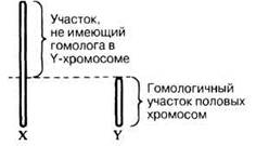
Fig. 24.20. Homologous and non-homologous regions of sex chromosomes.
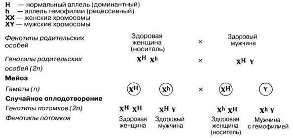
Fig. 24.21. Inheritance mechanism of the sex-linked hemophilia allele.
One of the best-documented Examples of hemophilia inheritance is the pedigree of the descendants of British Queen Victoria. It is believed that the hemophilia gene arose as a mutation in Queen Victoria herself or in one of her parents. Figure 24.22 shows how this gene was transmitted to her descendants.

Fig. 24.22. Inheritance of hemophilia by the descendants of Queen Victoria. The diagram includes only those descendants who were involved in transmitting hemophilia or were affected by the disease. The pedigree of the British royal family is extended to show why hemophilia has not appeared in any of Queen Victoria's descendants for seven generations.
24.11. Coat color in cats and body color in the magpie moth are controlled by a sex-linked gene (i.e., located on the X chromosome). In two experimental crosses, in which the homogametic sex in the parental generation was homozygous for this gene, the following results were obtained

Which sex is heterogametic in each of these species?
Last update: 06/08/2026
Editorial and Educational Adaptation: This material has been compiled based on the primary/original source text. The project team performed an editorial review, corrected technical inaccuracies, structured sections, and adapted the content for an educational format.
What was processed:
- elimination of formatting defects (OCR errors, structural breaks, corrupted characters);
- editorial organization of content;
- standardization of terminology in accordance with academic sources;
- verification of factual statements against the original source text.
All mentions of the author, publication year, and origin of the primary text have been preserved in accordance with the source.