BIOLOGY Volume 2 - A Guide to General Biology - 2004
14. TRANSPORT IN ANIMALS
14.5. Blood vessels
14.5.1. General structure
Blood circulating through the body passes through Arteries, capillaries, and Veins. In principle, the walls of both arteries and veins consist of three layers: 1) an inner layer, or endothelium (tunica intima), formed of squamous epithelium; 2) a middle layer (tunica media), consisting of smooth Muscle Cells and elastic fibers; and 3) an outer layer (tunica externa), composed primarily of inelastic Collagen fibers.
Structural Features of arteries and veins are listed in Table 14.3 and shown in Fig. 14.3. Some of the Tissues that make up these vessels are discussed in Chapter 6 (see Fig. 6.14, 6.22, and 6.23).
Class="center">Table 14.3. Structure AND Functions of arteries, capillaries, and veins (diagrams are not to scale)
Arteries |
Veins |
Capillaries |
Carry blood toward the heart1 |
Connect arteries and veins. Serve as the site of exchange of substances between blood and other tissues |
|
Middle layer thick, consisting of elastic fibers and smooth muscle cells |
Middle layer relatively thin, with few smooth muscle cells and elastic fibers |
Middle layer absent. The only tissue is squamous endothelium (sec. 6.3.1). Elastic fibers absent |
No internal Valves (except at the exit from the heart) |
Large veins have valves at regular intervals that prevent backflow of blood |
No internal valves |
Blood pressure high, pulsatile |
Blood pressure low, non-pulsatile |
Blood pressure decreasing, non-pulsatile |
Blood flows rapidly |
Blood flows slowly |
Blood flow slows down |
Blood volume small |
Blood volume much greater than in capillaries or arteries |
Blood volume large |
Blood is oxygenated, except in the pulmonary arteries |
Blood is deoxygenated, except in the Pulmonary veins |
Blood gradually becomes deoxygenated (the reverse occurs in the Lungs) |
|
|
|
1 Portal veins are an exception. |
||
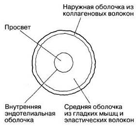
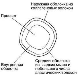
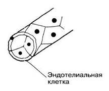
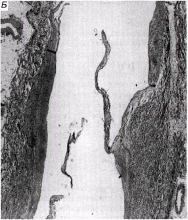
Fig. 14.3. A. Cross-section of an artery and a vein. B. Cytology/practical/54.html">Longitudinal section of a vein showing a valve.
Last update: 06/08/2026
Editorial and Educational Adaptation: This material has been compiled based on the primary/original source text. The project team performed an editorial review, corrected technical inaccuracies, structured sections, and adapted the content for an educational format.
What was processed:
- elimination of formatting defects (OCR errors, structural breaks, corrupted characters);
- editorial organization of content;
- standardization of terminology in accordance with academic sources;
- verification of factual statements against the original source text.
All mentions of the author, publication year, and origin of the primary text have been preserved in accordance with the source.