BIOCHEMISTRY - L. Stryer - 1984
VOLUME 2
PART III. BIOSYNTHESIS OF MACROMOLECULE PRECURSORS
CHAPTER 20. BIOSYNTHESIS OF MEMBRANE LIPIDS AND STEROID HORMONES
20.18. Absence of LDL Receptors Leads to Hypercholesterolemia and Premature Atherosclerosis
Studies on familial hypercholesterolemia highlight the crucial role of LDL receptors. In this disorder, the total concentration of Cholesterol and LDL in the Blood
serum is markedly elevated. This condition results from a mutation in a single autosomal locus. The serum cholesterol level is typically 6.8 g/L in homozygous patients, 3 g/L in heterozygotes, and 1.75 g/L in normal individuals. Because the concentration of cholesterol-bearing LDL in Blood Plasma is increased, cholesterol is deposited in various Tissues. These cholesterol deposits are called xanthomas and are frequently found in the Skin and tendons. More dangerous is the deposition of cholesterol in plaques along arterial walls, leading to atherosclerosis. Most homozygous hypercholesterolemic patients die in childhood due to CORONARY Heart DISEASE. Heterozygotes exhibit Various Forms of the condition, which generally follows a milder course. In most cases of familial hypercholesterolemia, the molecular defect consists of the absence or deficiency of functional LDL receptors. Homozygotes possess virtually none of these receptors, whereas heterozygotes have approximately half the normal amount. Consequently, the uptake of LDL by extrahepatic tissues is impaired, resulting in elevated serum LDL levels. Additionally, hereditary hypercholesterolemia may arise from a disruption in the internalization of the LDL-receptor complex.
Class="center">Fig. 20.17. Pathway of low-density lipoprotein METABOLISM in cultured human fibroblasts. HMG-CoA reductase stands for 3-hydroxy-3-methylglutaryl-CoA reductase; ACAT stands for acyl-CoA:cholesterol acyltransferase.

20.19. Steroid Nomenclature
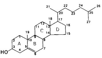
Before examining the synthesis of Steroid Hormones, we must introduce a few conventions regarding steroid nomenclature. The carbon atoms in Steroids are numbered as shown in Fig. 20.18, using cholesterol as an example. The rings of the steroid Nucleus are designated by the capital letters A, B, C, and D. Cholesterol contains two angular methyl groups: the C-19 methyl group is attached to C-10, and the C-18 methyl group is attached to C-13. Chemical bonds drawn pointing upwards from C-10 and C-13 denote these methyl groups. By definition, the C-18 and C-19 methyl groups of cholesterol lie above the plane of the four rings. Substituents located above this plane are referred to as β-oriented, and their bonds are represented by solid lines. Substituents located below the plane of the rings are in the α-orientation, and their bonds are depicted by dashed lines.
Fig. 20.18. Carbon atom numbering in the cholesterol molecule
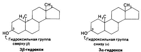
The hydrogen atom at the C-5 position can be in either the α- or β-orientation. When this hydrogen is α-oriented, rings A and B are fused in a trans-conformation, whereas a β-orientation results in a cis-fused ring system. When the stereochemistry at C-5 is unspecified, a trans-junction is assumed. In all steroid hormones that possess a hydrogen atom at C-5, it is in the α-orientation. Conversely, in Bile acid molecules, the C-5 hydrogen is in the β-orientation. Thus, cis-fusion is characteristic of bile acid salts, whereas trans-fusion is found in all steroid hormones bearing a hydrogen at C-5. Trans-fused rings yield an almost planar Structure, whereas cis-fusion confers a bent, curved shape.
20.20. Steroid Hormones Are Derived from Cholesterol
Cholesterol is the precursor of the five Major Classes of steroid hormones: progestins, glucocorticoids, mineralocorticoids, androgens, and estrogens (Fig. 20.19). Progesterone, one of the progestins, prepares the uterine lining for egg implantation and plays a vital role in maintaining Pregnancy. Androgens (such as testosterone) are responsible for The Development of male secondary sex characteristics, whereas estrogens (such as estradiol) are essential for female secondary sex characteristics and also participate in the ovarian cycle. Glucocorticoids (such as cortisol) stimulate Gluconeogenesis and Glycogen synthesis while promoting The breakdown of fats and Proteins. Mineralocorticoids (such as aldosterone) enhance the renal reabsorption of Na+, Cl-, and HSO3- ions, thereby increasing blood volume and blood pressure. These classes of hormones are synthesized primarily in specific tissues: progestins in the corpus luteum; estrogens in the Ovaries; androgens in the Testes; and glucocorticoids and mineralocorticoids in the adrenal cortex.
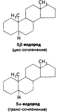
Fig. 20.19. Biosynthetic pathways of steroid hormones

20.21. Steroids Undergo Hydroxylation Catalyzed by NADPH- and Oxygen-Dependent Monooxygenases
Hydroxylation reactions play a critical role in the synthesis of cholesterol from squalene and in The conversion of cholesterol into steroid hormones and bile acids. All of these hydroxylations require NADPH and O2. Experiments utilizing O2 and H2O labeled with 18O have demonstrated that the oxygen atom of the introduced hydroxyl group is derived from O2 rather than from H2O. One atom of the O2 molecule is incorporated into the substrate, while the other is reduced to Water. Enzymes catalyzing such reactions are termed Monooxygenases (or mixed-function oxidases). Recall that another monooxygenase is also involved in the hydroxylation of phenylalanine (Section 18.16).
RH + O2 + NADPH + H+ → ROH + H2O + NADP+
Hydroxylation requires the activation of molecular oxygen. In The Biosynthesis of steroid hormones and bile acid salts, this activation is mediated by a specialized cytochrome designated as P450, so named because its carbon monoxide complex exhibits an absorption maximum at 450 nm. Cytochrome P450 is the terminal component of an electron-transport chain found in adrenal Cell/35.html">Mitochondria and Liver microsomes. The primary function of this chain is hydroxylation rather than Oxidative Phosphorylation. High-energy electrons from NADPH are transferred to a flavoprotein within the chain and subsequently passed to adrenodoxin, a non-heme iron protein. Adrenodoxin then delivers the electrons to the oxidized form of cytochrome P450. Finally, the reduced P450 activates O2.
The cytochrome P450 system plays a major role in the detoxification of foreign (xenobiotic) compounds. For instance, the hydroxylation of the barbiturate phenobarbital increases its water solubility and facilitates its excretion. Similarly, polycyclic aromatic Hydrocarbons are hydroxylated by the P450 system. The Introduction of hydroxyl groups enables the attachment of highly polar moieties (such as glucuronate or sulfate), which markedly enhance the aqueous solubility of these modified aromatic molecules. However, the action of the P450 system is not always beneficial to the Organism. Recent research has shown that many potent carcinogens are converted in vivo into chemically reactive forms, a metabolic activation process typically carried out by the P450 system.
Last update: 06/08/2026
Editorial and Educational Adaptation: This material has been compiled based on the primary/original source text. The project team performed an editorial review, corrected technical inaccuracies, structured sections, and adapted the content for an educational format.
What was processed:
- elimination of formatting defects (OCR errors, structural breaks, corrupted characters);
- editorial organization of content;
- standardization of terminology in accordance with academic sources;
- verification of factual statements against the original source text.
All mentions of the author, publication year, and origin of the primary text have been preserved in accordance with the source.