BIOCHEMISTRY - L. Stryer - 1984
VOLUME 1
PART I. CONFORMATION AND DYNAMICS
CHAPTER 9. CONNECTIVE TISSUE PROTEINS: COLLAGEN, ELASTIN, AND PROTEOGLYCANS
9.10. Extension peptides of precursor chains are enzymatically cleaved
The pro-α1 (I) and pro-α2 chains are synthesized by fibroblasts and secreted into the extracellular space of Connective Tissue. Here, the extension Peptides at the N- and C-termini of these precursor chains are cleaved by specific Proteolytic Enzymes, procollagen peptidases.
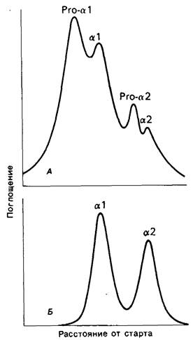
Impaired conversion of precursor chains to α1 and α2 chains leads to generalized connective tissue damage. For example, patients with Ehlers-Danlos syndrome are characterized by short stature, hyperextensible Skin, and hypermobile joints. In one form of this disease, significant amounts of procollagens are found in extracts of patients' skin and tendons (Fig. 9.14). Concurrently, a decrease in procollagen peptidase activity is detected in fibroblasts. There is also an inherited (recessive) disease in cattle called dermatosparaxis, which is caused by a deficiency of procollagen peptidase. The skin of these animals becomes extremely fragile because the Collagen fibers are present as disorganized bundles. Furthermore, a significant portion of the existing mature type I collagen contains α1 (I) and α2 chains with retained N-terminal procollagen peptides. All of this indicates that the Cleavage of N-terminal peptides is essential for The formation of ordered collagen fibrils.
Class="center">Fig. 9.14. Polyacrylamide gel Electrophoresis of skin extracts. Acid extracts of skin from patients with Ehlers-Danlos syndrome type VII (A) contain a significant amount of procollagen (pro-α1 and pro-α2). In normal controls (B), procollagen is absent
9.11. Collagen fibers consist of staggered tropocollagen molecules
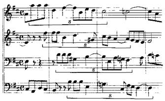
The formation of collagen fibers occurs in the extracellular space As a result of the spontaneous, specific self-assembly of tropocollagen molecules. The Structure of collagen fibers has been studied by X-Ray Diffraction and Electron Cell/15.html">Microscopy. Collagen fibers exhibit a transverse striation pattern with a periodicity of 680 Å (see Fig. 9.1). In contrast, the length of a tropocollagen molecule is 3000 Å. Thus, the structural period of the fiber is several times shorter than the length of its constituent molecules; this indicates that the rows of tropocollagen molecules cannot lie exactly end-to-end. Indeed, one row of tropocollagen molecules is staggered relative to the adjacent row by approximately 1/4 of the molecular length. It follows that the Structural Organization of the collagen fiber is based on parallel rows of tropocollagen molecules staggered by a quarter-period (Fig. 9.15). This "arrangement" resembles a musical fugue (Fig. 9.16).
Fig. 9.15. Schematic representation of the structural organization of a collagen fiber. Tropocollagen molecules (indicated by blue arrows) are arranged in rows, sequentially staggered by 1/4 relative to one another. The gaps between tropocollagen molecules in the rows (indicated by red squares) can serve as nucleation sites for calcification during Bone Formation
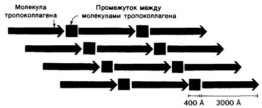
Fig. 9.16. Passage from the Fugue in D major, "The Well-Tempered Clavier" by J. S. Bach
An interesting structural feature of the fiber is that the tropocollagen molecules aligned in a row are not connected "end-to-end." There is a gap of about 400 Å between the end of one molecule and the beginning of the next (Fig. 9.15). This gap plays a special role in bone formation. Bone consists of an organic phase, almost entirely represented by collagen, and an inorganic phase, namely calcium phosphate. The latter is structurally close to hydroxyapatite, with the composition Ca10 (PO4)6 (OH)2. Collagen is essential for bone formation as a site for the deposition of calcium phosphate crystals. It turns out that the first crystals are deposited at intervals of about 680 Å, which coincides with the periodicity of the collagen fiber. It is highly likely that the gaps along the rows of tropocollagen molecules serve as nucleation sites for the deposition of the mineral components of bone.
9.12. Collagen fiber formation is regulated by procollagen peptidases
Unlike tropocollagen, procollagen molecules are incapable of spontaneous fiber formation; this requires the prior Cleavage of the N- and C-terminal peptides. Thus, the formation of collagen fibers is analogous to the assembly of fibrin threads. Procollagen is analogous to fibrinogen, tropocollagen to fibrin monomers, and procollagen peptidases to Thrombin. In both systems, specific proteolytic cleavage is a prerequisite for fiber formation.
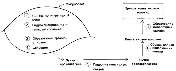
Fibroblasts secrete procollagen rather than tropocollagen (Fig. 9.17). The mechanisms of Biosynthesis and secretion are analogous to those operating for pancreatic zymogens (Section 8.1) and other secretory Proteins (Section 29.29). The collagen fiber is formed in the extracellular fluid near The surface of the fibroblast, not inside it, because procollagen peptidases are located extracellularly. The terminal Peptides of the precursor chains prevent premature fiber formation. They may also be involved in The transport of procollagen across the fibroblast membrane. At an earlier intracellular stage, these extension peptides facilitate the alignment and association of the three chains for subsequent triple helix formation. Interchain Disulfide Bonds are of particular importance in this process.
Fig. 9.17. Stages in the formation of a mature collagen fiber
9.13. Cross-links increase the strength of collagen fibers
Like fibrin, collagen is stabilized by covalent cross-links. Collagen fibers feature Two Types of cross-links: intramolecular (within a single tropocollagen unit) and intermolecular (between individual tropocollagen units). These cross-links are found only in two closely related proteins, Collagen and Elastin.
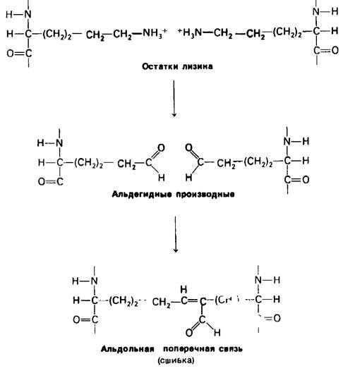
The side chains of Lysine participate in the formation of intramolecular cross-links in collagen. In the first step, the ε-amino group of lysine is converted into an aldehyde by the enzyme lysyl oxidase. Next, two aldehydes undergo an aldol Condensation reaction (Fig. 9.18). In this reaction, the enolate ion formed from one aldehyde adds to the carbonyl group of the other aldehyde. The localization of this intramolecular cross-link is interesting. In the Amino Acid Sequence of the amino-terminal region of the α1 (I) collagen chain, Glycine becomes every third residue only starting from position 13. Consequently, lysine-5 is located in the non-helical region of the polypeptide chain, near the beginning of the triple helix. The flexibility of this non-helical segment facilitates the Formation of the cross-link in question.
Fig. 9.18. Formation of an aldol cross-link from two lysine side chains
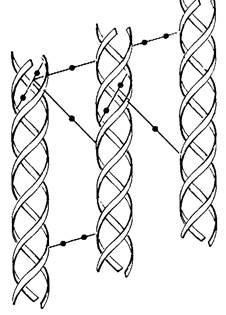
An aldol cross-link can be converted, through interaction with a Histidine side chain, into a histidinoaldol cross-link (Fig. 9.19). The aldehyde group of this aldol-histidine complex can form a Schiff base with another side chain, such as a hydroxylysine side chain. This can result in the covalent linking of four side chains to one another, some of which may belong to different tropocollagen molecules (Fig. 9.20).
Fig. 9.19. A histidine side chain can add across the C=C double bond of the aldol cross-link, forming a histidinoaldol cross-link
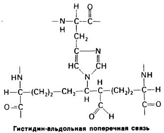
Fig. 9.20. Intra- and intermolecular cross-links in collagen
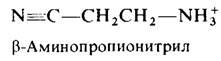
The number and type of cross-links in collagen depend on the function and age of the tissue. For instance, rat Achilles tendon collagen is characterized by a high degree of cross-linking, whereas the collagen of the more elastic tail tendon contains far fewer cross-links. The Significance of cross-links in providing collagen fibers with high tensile strength is clearly demonstrated in lathyrism, a disease in animals caused by eating the seeds of the sweet pea Lathyrus odoratus. The toxic agent in this case is β-aminopropionitrile, which inhibits The conversion of the lysine side chain into an aldehyde. Under METABOLISM/18.html">The Influence of this toxin, the collagen of affected animals loses its mechanical strength.
Last update: 06/08/2026
Editorial and Educational Adaptation: This material has been compiled based on the primary/original source text. The project team performed an editorial review, corrected technical inaccuracies, structured sections, and adapted the content for an educational format.
What was processed:
- elimination of formatting defects (OCR errors, structural breaks, corrupted characters);
- editorial organization of content;
- standardization of terminology in accordance with academic sources;
- verification of factual statements against the original source text.
All mentions of the author, publication year, and origin of the primary text have been preserved in accordance with the source.