BIOCHEMISTRY - L. Stryer - 1984
VOLUME 1
PART I. CONFORMATION AND DYNAMICS
CHAPTER 9. CONNECTIVE TISSUE PROTEINS: COLLAGEN, ELASTIN, AND PROTEOGLYCANS
9.8. Impaired Hydroxylation Is a Biochemical Defect in Scurvy
Structure/19.html">The Importance of Collagen hydroxylation is vividly illustrated by scurvy. In 1536, Jacques Cartier (J. Cartier) gave a graphic description of this disease, which afflicted his companions during their voyage up the St. Lawrence River:
"Some lost all their strength and could not stand on their feet... Others also had purple spots of Blood on their Skin, which gradually covered their shins, knees, thighs, buttocks, shoulders, arms, and neck. Their mouths became stinking, their Gums so rotten that all the flesh fell off to the roots of the Teeth, and almost all their teeth fell out."
The means of preventing scurvy were concisely formulated by the English physician James Lind (J. Lind), who wrote in 1753:
"Experience has repeatedly shown that greens, fresh vegetables, and ripe fruits are not only the best remedy, but also the most effective means of preventing this disease."
Lind urged that lemon juice be included in the sailors' diet. The British Admiralty acted on his advice about 40 years later.
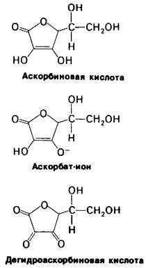
Scurvy is caused by a deficiency of ascorbic acid (Vitamin C) in the diet. Primates and guinea pigs have lost The ability to synthesize ascorbic acid and must therefore obtain it from food. As a strong reducing agent, ascorbic acid (Fig. 9.12) prevents the inactivation of prolyl hydroxylase, apparently by maintaining the iron atom in the enzyme in its reduced state. Collagen synthesized in the absence of ascorbic acid is underhydroxylated and, consequently, has a lower melting Temperature. Such collagen cannot form structurally normal fibers, which leads to the skin lesions and blood vessel fragility so characteristic of scurvy.
Class="center">Fig. 9.12. Structures of ascorbic acid (vitamin C) and its ionized form, ascorbate. The pKa of the acidic hydroxyl groups of ascorbic acid is 4.2. Dehydroascorbic acid is the oxidized form of the ascorbate ion
9.9. Procollagen Is a Precursor of Collagen in Its Biosynthesis
The triple helix of type I collagen forms very rapidly in vivo. In vitro, however, the assembly of the triple helix from dissolved α1(I) and α2 chains takes days and has a low yield. What accounts for this difference in tropocollagen helix assembly in vivo and in vitro? For comparison, recall how The structure of denatured chymotrypsinogen and Chymotrypsin is restored: the denatured proenzyme spontaneously assumes its correct three-dimensional conformation, whereas the active enzyme cannot. This is because chymotrypsin lacks a portion of the structure required for proper folding—namely, two dipeptides that are cleaved during activation.
By analogy, one might expect that the inability of purified α1(I) and α2 chains to spontaneously form the correct tropocollagen structure is due to the absence of certain elements carrying the necessary information. Indeed, this is the case. The constituent chains of collagen are synthesized as larger precursor molecules. The precursor of the α1(I) chain, called pro-α1(I), has a mass of 140 kDa (whereas the α1(I) chain has a mass of 95 kDa). Additional Peptides are located at both the N- and C-termini of the pro-α1(I) chain (Fig. 9.13). Similarly, the precursor of the α2 chain is pro-α2, with a mass of 140 kDa. In terms of Amino Acid Composition, the additional peptide regions in pro-α1(I) and pro-α2 are completely unlike the main body of the chain. They contain little Glycine, Proline, and hydroxyproline. The N-terminal peptides in pro-α1(I) and pro-α2 contain intrachain Disulfide Bonds. Furthermore, the C-terminal peptides of these precursor chains are linked by interchain disulfide bonds, which are absent in mature collagen.
Fig. 9.13. Schematic representation of The conversion of procollagen to collagen by Cleavage of the N-terminal peptide (about 15 kDa) and the C-terminal peptide (about 30 kDa) from each of the three chains. In procollagen, the C-terminal Peptides of the three chains are linked by disulfide bridges

Last update: 06/08/2026
Editorial and Educational Adaptation: This material has been compiled based on the primary/original source text. The project team performed an editorial review, corrected technical inaccuracies, structured sections, and adapted the content for an educational format.
What was processed:
- elimination of formatting defects (OCR errors, structural breaks, corrupted characters);
- editorial organization of content;
- standardization of terminology in accordance with academic sources;
- verification of factual statements against the original source text.
All mentions of the author, publication year, and origin of the primary text have been preserved in accordance with the source.