BIOCHEMISTRY - L. Stryer - 1984
VOLUME 1
PART I. CONFORMATION AND DYNAMICS
CHAPTER 6. INTRODUCTION TO ENZYMOLOGY
6.21. The Binding of Substrates to Enzymes Involves Highly Oriented Hydrogen Bonds
Although many substrates are uncharged, they bind to Enzymes with a high degree of Specificity and affinity. The primary mode of interaction for such substrates, as well as for most charged substrates, with enzymes is The formation of Hydrogen Bonds. In a Hydrogen bond, a hydrogen atom is shared between two other atoms. The atom to which the hydrogen is more firmly bound is called the hydrogen donor, whereas the second atom is the hydrogen acceptor. Essentially, a hydrogen bond can be viewed as an intermediate interaction that occurs during The transfer of a proton from an acid to a base. The acceptor atom must possess a partial negative charge, which attracts the hydrogen. In short, a hydrogen bond is somewhat reminiscent of a ménage à trois:
Class="center">
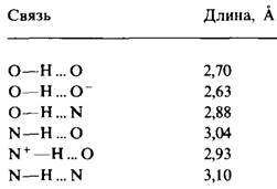
In biological systems, the donor atoms in Hydrogen bonds are typically nitrogen or oxygen atoms covalently bonded to a hydrogen atom. Oxygen or nitrogen acts as the acceptor atoms. The types of Hydrogen bonds and their lengths are listed in Table 6.3. Bond energies range from approximately 3 to 7 kcal/mol. Hydrogen bonds are stronger than Structure/103.html">Van der Waals interactions, but significantly weaker than covalent bonds. In terms of length, hydrogen bonds occupy an intermediate position between covalent bonds and van der Waals interactions. An important feature of hydrogen bonds is that their energy depends on geometry. A hydrogen bond is strongest when the donor, hydrogen, and acceptor lie in a straight line. If the acceptor atom is positioned at an angle relative to the line connecting the donor atom and the hydrogen, the bond becomes weaker as this angle increases:

Table 6.3. Typical dimensions of hydrogen bonds
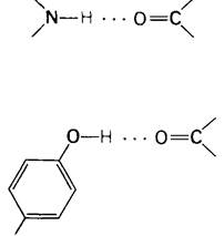
We have already mentioned hydrogen bonds when discussing The structure of Myoglobin and Hemoglobin. In an α-Helix, a hydrogen bond connects the —NН— and —СО— groups of the peptide chain. Here, the nitrogen atom acts as the hydrogen donor and the oxygen atom as the acceptor. The distance between the nitrogen and oxygen atoms is 2.9 Å; the hydrogen atom is 0.9 Å closer to the nitrogen than to the oxygen.
Another example of a hydrogen bond in myoglobin and hemoglobin is the interaction between the hydroxyl group of Tyrosine HC2 and the peptide carbonyl FG4. The oxygen atom of the tyrosine hydroxyl group serves as the hydrogen donor, and the oxygen atom of the peptide C=O group serves as the acceptor:
The Role of hydrogen bonds in enzyme-substrate interactions is clearly illustrated by the binding of the uridine-containing portion of the substrate to pancreatic Ribonuclease, an enzyme that cleaves ribonucleic acid (Fig. 6.25). In this case, three hydrogen bonds are formed.
Fig. 6.25. Hydrogen bond formation during the binding of a substrate to ribonuclease

1. One of the C—O groups of the uridine ring is hydrogen-bonded to the N—H group of the peptide backbone.
2. The N—H group of the uridine ring is hydrogen-bonded to the —OH group of a Threonine residue.
3. Another C=O group of the uridine ring is hydrogen-bonded to the —OH group of a Serine residue.
6.22. Proteins Have a High Capacity for Hydrogen Bonding
Amino acid side chains and the main peptide backbone can form A large number of diverse hydrogen bonds. For instance, of the 20 Amino Acids found in Proteins, 11 have side chains capable of participating in hydrogen bonding. It is convenient to classify these amino acids into groups based on the types of hydrogen bonds they form.
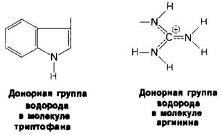
1. The side chains of Tryptophan and Arginine can function exclusively as hydrogen Donors:
2. The side chains of asparagine, glutamine, serine, and threonine can act as both hydrogen donors and acceptors, making them similar in this respect to the peptide group.
3. The ability to form hydrogen bonds in Lysine (and generally in compounds with terminal amino groups), in aspartic and glutamic acids (and Other Compounds with terminal carboxyl groups), as well as in pyridine and Histidine, is pH-dependent. Within certain pH ranges, they can act simultaneously as both hydrogen donors and acceptors, whereas at other pH values they function either as donors or as acceptors, as illustrated for aspartate and glutamate in Fig. 6.26. In other words, the type of hydrogen bond formed by these ionizable residues depends on the pH.
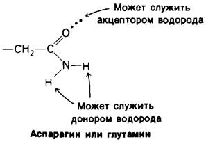
Fig. 6.26. Hydrogen-bonding capacity of aspartate and glutamate

Last update: 06/08/2026
Editorial and Educational Adaptation: This material has been compiled based on the primary/original source text. The project team performed an editorial review, corrected technical inaccuracies, structured sections, and adapted the content for an educational format.
What was processed:
- elimination of formatting defects (OCR errors, structural breaks, corrupted characters);
- editorial organization of content;
- standardization of terminology in accordance with academic sources;
- verification of factual statements against the original source text.
All mentions of the author, publication year, and origin of the primary text have been preserved in accordance with the source.