Biochemistry - The Chemical Reactions of Living Cells Volume 3 - D. Metzler 1980
Biochemical Genetics and the Synthesis of Nucleic Acids and Proteins
Recombination, Integration, and Excision
Mechanisms of Recombination
Let us now briefly examine the somewhat obscure chemical phenomena underlying such processes as genetic recombination, the integration of viral DNA into the host Cell genome, and the excision of a prophage from the host cell chromosome. The complexity of the recombination process is evidenced by the fact that in mutants defective in recombination ability, Mutations are localized not in a single region (Gene) of the E. coli chromosome, but in several; the corresponding genes are designated as rесА, В, С, F, G, and Н. Bacteria with mutations in some of these genes are exceptionally sensitive to ultraviolet irradiation, indicating their inability to repair (restore) DNA Damage caused by UV light (Ch. 13, Sec. D, 2). It follows that some of the Enzymes required for recombination are also needed by The Cell to repair UV-induced damage. However, the specific Functions of most of these gene products remain incompletely understood. It is believed that E. coli possesses two fully functional general recombination systems. The λ phage genome contains genes encoding another recombination system that functions independently of the λ phage inf and xis gene products (Fig. 15-15), which are necessary for the integration and excision of viral genetic material and mediate Site-Specific Recombination (for specific Regions of the genomes) between the host and viral genes.
The most puzzling aspect of recombination is how homologous segments of two different double-stranded DNA molecules become joined. As shown schematically in equation (15-10), the exchange of polynucleotide chain segments must occur at precisely the same point in each of the two double-stranded molecules.
Class="center">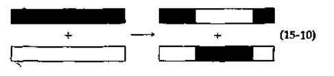
One of the earliest mechanisms proposed to explain genetic recombination was based on the assumption that recombination is directly coupled to DNA Synthesis. According to this "copy-choice" mechanism, Replication proceeds along One DNA strand up to some random point, at which the polymerase jumps to the second of the two homologous Chromosomes and begins copying it. Consequently, the newly formed DNA molecule would be partially complementary to one parental double-stranded DNA molecule and partially to the other. To test this hypothesis, Meselson and Weigle [220] infected E. coli with two λ phage strains containing DNA labeled with stable isotopes of carbon (13С) and nitrogen (15N), respectively. Density gradient centrifugation revealed that the recombinant DNA contained both 13С and 15N. Thus, it became clear that the recombinant progeny DNA incorporates DNA from both parents. This result disproved the copy-choice hypothesis and supported a mechanism in which recombination is accompanied by strand breakage.
If recombination occurs via the Enzymatic Cleavage of two homologous double-stranded DNA molecules (followed by rejoining), the question arises: how does the cell avoid gene inactivation due to the addition or loss of genetic material? It seems implausible that recombination could occur through the random action of nonspecific enzymes and random rejoinings. At the same time, experience shows that general recombination can occur at any point in The Genome with a fairly constant frequency along the entire length of the DNA chain. Clearly, these facts can only be understood in terms of complementary base pairing between homologous regions of single strands from two different double-stranded DNA molecules.
One recombination model was based on data derived from The Study of phages λ and T4. According to this model, the λ phage exo gene (Fig. 15-22) is not required for replication but is essential for general recombination. The product of this gene has been shown to be a 5'-3' exonuclease. A possible MECHANISM OF ACTION for this enzyme during recombination is illustrated in Fig. 15-31. The process begins with an endonuclease introducing single-strand breaks at random sites in the double-stranded DNA molecules. A specialized exonuclease then widens these breaks, converting them into gaps. The exposed homologous regions of one molecule will tend to associate with complementary regions of another molecule (Fig. 15-31, stage b), forming H-shaped heteroduplex structures. Branch migration (Fig. 15-31, stage c) leads to the elongation of the heteroduplex region and the appearance of a short branch. In the case of replicating phage T4, electron micrographs [221] of branched DNA molecules of the type shown in Fig. 15-29 have been obtained. Endonuclease action on these branched structures (Fig. 15-31, stage d) will produce "nicks." Any single-stranded gaps can be filled by DNA polymerase (Fig. 15-31, stage c), and the breaks can be sealed by polynucleotide ligase.

FIG. 15-31. A possible mechanism of recombination involving The formation of point breaks at random sites in homologous double DNA strands, the widening of these breaks into gaps, and the association of complementary regions. The branch migration mechanism subsequently fills the resulting gaps and reseals all nicks, yielding a recombinant DNA molecule.
An argument against this recombination mechanism is that it requires the formation of rather long single-stranded tails in the DNA molecules. In view of this, Holliday [222] proposed a model that does not require such tails. The recombination process can begin at specific sites on double-stranded DNA molecules recognized by a recombination enzyme (Fig. 15-32, A). After localized unwinding of the DNA, an exchange takes place between the two broken strands, which are then rejoined by ligase as shown in Fig. 15-32, A. As both helices rotate about their axes, the crossover points migrate up or down the chains, leading to the formation of heteroduplex DNA regions. The ability of the process to halt at any distance from the initiation point helps explain the universality of genetic recombination. The process concludes with strand cleavage and the rejoining of the two strands. If the strands originally broken during initiation are rejoined (cleavage at points aa' in Fig. 15-32, A), genes located outside the heteroduplex region will not recombine, whereas Cleavage of the other strands (points bb') will result in recombination of these genes. The intermediates predicted by the Holliday model have been directly observed using the Electron microscope (Fig. 15-32, B) [222a].
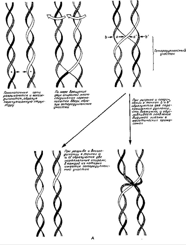
FIG. 15-32. A. Holliday recombination mechanism involving single-strand exchanges.
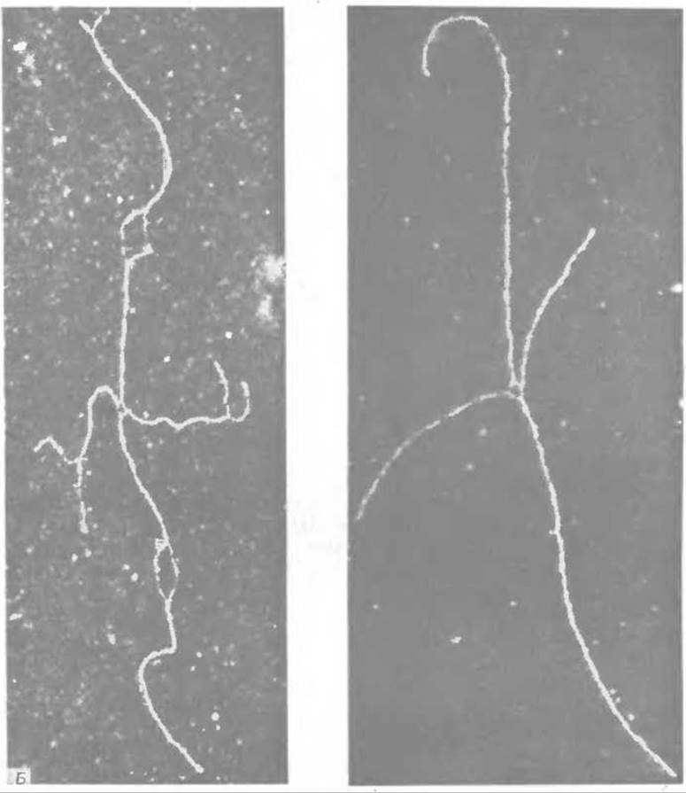
B. "χ-form" of colicin E1 plasmid DNA. These forms are thought to arise from figure-eight-shaped Holliday intermediates whose length is twice that of the colicin genome. These figure-eights are cleaved by the restriction enzyme Eco RI (Sec. E, 1) at a specific site that occurs only once in the genome, yielding "y-forms." The pairs of short and long arms are believed to represent homologous duplexes. At the crossover point, the single strands can be seen to diverge, clearly demonstrating the strand junctions. This Structure corresponds to the intermediate predicted by the Holliday model (top right in Fig. A); it can be formed, for example, by the rotation of one vertical duplex around the other. The DNA specimen shown on the right was prepared at high formamide concentrations, allowing visualization of Denaturation loops and "frayed" ends in AT-rich regions (Sec. D, 3) [222a].
Such cross-stranded structures can be formed by any two intact double-stranded DNA molecules of arbitrary nucleotide composition [223, 224]. All that is required is the Introduction of nicks into each polynucleotide chain and the rejoining of non-identical strands to bridge the gap between the double-stranded molecules. Heteroduplex formation can propagate along the chains via mutual rotation. This model also explains why the crossed strands are cleaved precisely at the points required to complete DNA recombination.
Several modifications of the model presented in Fig. 15-32 have been proposed [223, 225]. According to the model of Sobell [226, 227] and others [228], initial nicks occur within cruciform structures formed at palindromic sequences (Fig. 15-4).
During the recombination of phages S13 and φX174, circular dimers serve as intermediates [229]. Such double-length rings, as well as catenated (interlocked) rings of normal length, can be generated by recombination proceeding via the strand-breakage mechanisms discussed in previous sections.
Our understanding of recombination is still far from complete, and some observations remain difficult to interpret. For instance, the recB and recC genes of E. coli encode Proteins that associate to form an unusual DNA-hydrolyzing enzyme [230]. Among its other functions, this enzyme, which has a total Molecular Weight of approximately 340,000, is capable of endonucleolytically cleaving single-stranded DNA. Although it cannot nick intact double-stranded DNA molecules, it may participate in generating putative endo- and exonucleolytic breaks during recombination. Interestingly, this enzyme requires ATP for its activity. In vitro, for every phosphodiester bond of DNA cleaved, about 20 molecules of ATP are hydrolyzed to ADP and Pi. In the absence of the DNA substrate, no ATP cleavage occurs. While one may doubt whether this enzyme functions in vitro exactly as it does in vivo, its intriguing properties and its necessity for recombination make it an important subject for future research. According to available data, a similar enzyme isolated from Bacillus subtilis joins double-stranded DNA molecules to one another (as shown by Electron Microscopy) [231]. The Significance of this effect remains unclear.
Recombination in Eukaryotic Cells occurs predominantly during Meiosis within the synaptonemal complex—a structure located between a pair of homologous chromatids. It has been suggested that the "nodules" periodically observed within this complex may be related to the recombination process [232]. In any case, it is clear that Crossing-over and recombination in Eukaryotic cells are complex, highly organized, and inextricably linked processes about which we currently know very little.
Last update: 06/08/2026
Editorial and Educational Adaptation: This material has been compiled based on the primary/original source text. The project team performed an editorial review, corrected technical inaccuracies, structured sections, and adapted the content for an educational format.
What was processed:
- elimination of formatting defects (OCR errors, structural breaks, corrupted characters);
- editorial organization of content;
- standardization of terminology in accordance with academic sources;
- verification of factual statements against the original source text.
All mentions of the author, publication year, and origin of the primary text have been preserved in accordance with the source.