Biochemistry - The Chemical Reactions of Living Cells, Volume 1 - D. Metzler 1980
The Molecules We Are Made Of
Sugars and Polysaccharides
Conformation of Polysaccharide Chains
Despite the great variety of monomeric units and types of linkages between them, the conformational flexibility of carbohydrate chains is quite limited. The sugar ring represents a rigid structural unit, and the joining of two rings can be described by two torsion angles, φ and ψ, much like the approach used for Peptides [48, 49]. Admittedly, there is no strict consensus on which polysaccharide conformation should correspond to φ and ψ values of 0°. Generally speaking, There is a rough empirical rule of this kind applicable to all polymers [18], but it is highly inconvenient when dealing with Polysaccharides. It is probably best to assume that φ and ψ are equal to 0° when the planes bisecting the sugar rings are coplanar:
Class="center">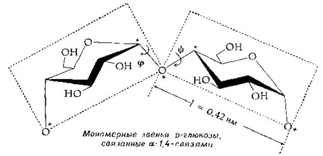
These planes pass through the atoms marked with asterisks in the figure and are generally not perpendicular to the plane of the rings. A systematic Study of the possible values of φ and ψ for Cellulose (poly-β-D-glucose) shows that these angles are confined to a very narrow range, in which the arrangement of monomeric units corresponds to an almost fully extended conformation.

Each glucose unit is rotated by 180° relative to the preceding one. The polymer possesses a twofold screw axis, with the ring planes exhibiting a slight zigzag pattern [50, 51]. Recall that in the chair conformation of glucose, all OH groups lie in the equatorial plane and are capable of forming Hydrogen Bonds with neighboring chains. This feature, combined with conformational rigidity due to the β-configuration of the monomeric units, undoubtedly accounts for the ability of cellulose to form strong fibers.
In starch and Glycogen, the chain is also formed by glucose residues, but in this case via an α-1,4-linkage. An extended conformation is no longer possible, and the chains coil into a helix. Among the Helical structures formed by Biopolymers, one of the first to be discovered (in 1943) [52] was the left-handed amylose helix winding around iodine (I2) molecules in the well-known iodine-starch complex (Fig. 2-17). The number of residues per turn is 6, the pitch of the helix is 0.8 nm, and the diameter is about 14 nm [53, 54].
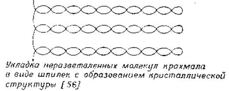
Another Structure—a more tightly wound double helix—has been proposed for amylose [55]. It is assumed that in such a helix each chain contains 6 residues per turn, and both chains run either in the same or in opposite directions. The amylose molecule contains an average of 1000 glucose residues and can be extended into a thin chain 500 nm in length, which exceeds the dimensions of crystalline regions in starch granules. Consequently, the chains within the granules must fold in some manner (for instance, into hairpin-like loops):
Agarose (mol. wt. 120,000) is a carbohydrate polymer with an alternating sequence of units.
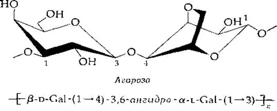
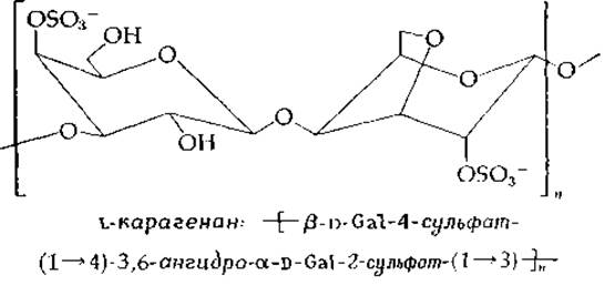
It is the major component of Agar and largely determines the remarkable gel-forming ability of this substance. It is possible to form a solid agar gel containing 99.5% Water. The molecular structure of agarose is based on a left-handed double helix with a threefold screw axis; the pitch of the helix is 1.90 nm, and its inner cavity is filled with water [57]. Gel-forming carrageenans from red marine Algae have a similar structure. According to X-ray data, in this case, three disaccharide units form a single turn of a right-handed helix with a pitch of 2.6 nm. The second chain runs parallel but is shifted by a half-turn, winding around the first helix [58]. At the intersection points of the double-helical regions, "junction zones" are formed, which act as gelation centers [59] (Fig. 2-18). Sulfate groups project laterally in pairs, serving as binding sites for Calcium Ions that stabilize the gel.
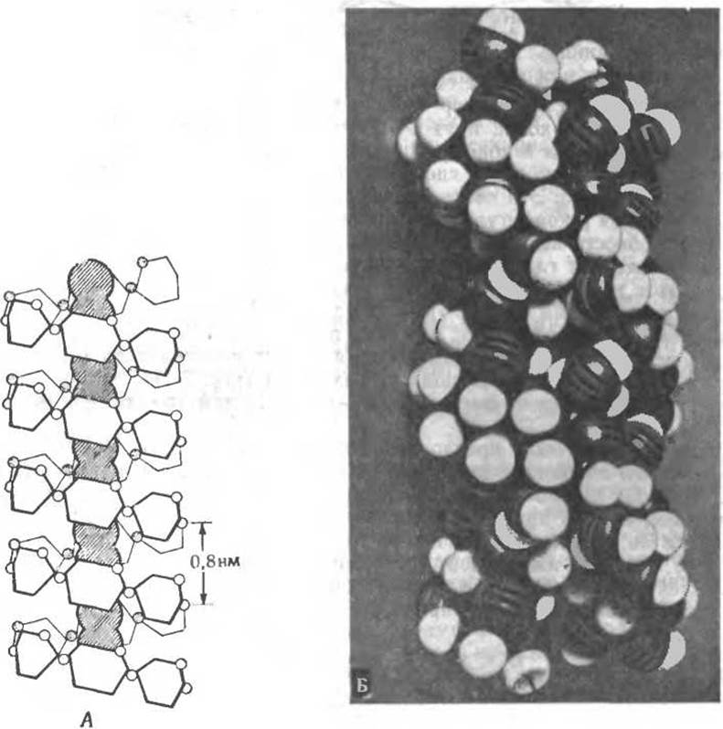
FIG. 2-17. A. STRUCTURE OF THE helical complex of amylose with iodine (I2). Iodine molecules are located along the axis of the helix formed by glucose residues, with six residues per turn. B. Model of a double helix composed of two parallel chains. Each chain contains 6 glucose residues per turn. According to the model, the periodicity is 2.1 nm (measured every 6 glucose residues along either chain). (Courtesy of D. French.)

FIG. 2-18. Schematic representation of the network structure of an agarose gel (right) compared with a random-coil network shown for reference (such a network is characteristic of Sephadex, among others). Agarose gels contain aggregates of 10 to 104 helices; smaller numbers of helices, as shown here, are typically not observed [58].
The occasional appearance of excess sulfate groups in these polymers introduces kinks into the chain (since the modified pyranose rings adopt a different type of chair conformation). These kinks prevent the polysaccharide from forming a single regular helix [59a].
The Xylan of higher plants, a β-1,3-polymer of D-xylose, is apparently a three-stranded right-handed triple helix [60]. Hyaluronic acid is hypothesized to have a double-helical structure [61]. Chondroitin sulfates consist of various types of single helices [62].
Supplement 2-B
Silicon: An Essential Trace Element
It is well known that active silicon METABOLISM takes place in the Cells of diatoms, whose skeletons are built of SiO2. Radiolarians, certain higher plants, and Sponges are also capable of accumulating silicon; in limpets (a type of mollusk), it forms The basis of the radular Teeth. Despite all these facts, silicon metabolism had not attracted much attention from researchers until it was recently demonstrated that this element is vital for the GROWTH AND DEVELOPMENT of higher animalsb-d. Specifically, in chicks, silicon has been found in sites of active ossification in developing bonesg. In the Internal Organs of mammals, silicon is present only in small quantities, yet in the Skin, Cartilage, and ligaments, its content reaches ~0.01%. Schwarz3 established that silicon is a constituent of mucopolysaccharides, such as chondroitin 4-sulfate, dermatan sulfate, and heparan sulfate. All of them contain ~0.04% silicon, meaning there is one atom of this element per 130–280 repeating polysaccharide units. In the plant kingdom, the silicon content in Pectins is approximately five times higher. Silicon in polysaccharides is very tightly bound, presumably via an ester linkage. Schwarz suggested that orthosilicic acid Si(OH)4 reacts with the hydroxyl groups of CARBOHYDRATES to form ester bonds, which may act as bridges between chains:

It should be noted that each of these structures contains unsubstituted OH groups at the silicon atom, As a result of which cross-links can form simultaneously between several polysaccharide chains. Based on these findings—which are admittedly preliminary—it can be hypothesized that the Biological Role of silicon in Connective Tissue lies in its ability to facilitate cross-linking (see also Chapter 11, Section D.3).
a Schwarz K., PNAS, 70, 1608–1612 (1973).
b Schwarz K. In: Trace Element Metabolism in Animals (Mills F., ed.), pp. 25–38, Livingstone, Edinburgh, 1970.
в Schwarz К., Milne D. B., Nature (London), 239, 333—334 (1972).
r Carlisle E. M., Science, 178, 619—621 (1972).
д Hoekstra W. H., Suttie J. W., Ganther H. E., Mertz W. eds., Trace Element Metabolism in Animals — 2, Univ. Park Press, Baltimore, Maryland, 1974.
Last update: 06/08/2026
Editorial and Educational Adaptation: This material has been compiled based on the primary/original source text. The project team performed an editorial review, corrected technical inaccuracies, structured sections, and adapted the content for an educational format.
What was processed:
- elimination of formatting defects (OCR errors, structural breaks, corrupted characters);
- editorial organization of content;
- standardization of terminology in accordance with academic sources;
- verification of factual statements against the original source text.
All mentions of the author, publication year, and origin of the primary text have been preserved in accordance with the source.