Human Biochemistry, Volume 1 - Murray R. 1993
Structure and Functions of Proteins and Enzymes
Enzymes: General Properties
Isoenzymes (Isozymes)
When we use terms such as "malate dehydrogenase" or "glucose-6-phosphatase", we usually refer to a specific protein with enzymatic activity. However, these names actually encompass all Proteins that catalyze The oxidation of malate to oxaloacetate or the Hydrolysis of glucose-6-phosphatase to yield glucose and Pi. Specifically, following the isolation of malate dehydrogenase from various sources (such as rat Liver and E. coli), it was found that the Enzymes from these different sources catalyzing the same reaction differ considerably in their Physical and Chemical properties. Physically distinct forms of enzymes possessing the same type of catalytic activity can occur in different Tissues of the same Organism, in different Cell types of the same tissue, and even within a prokaryotic organism such as E. coli. This discovery was made possible by the application of protein electrophoretic Separation Methods, which revealed electrophoretically distinct forms of a given enzymatic activity.
The term "isoenzyme" (or "isozyme") covers all the aforementioned physically distinct proteins with a given catalytic activity. In practice, however—particularly in clinical medicine—it is used in a narrower sense to denote physically distinct and separable forms of a given enzyme present in different cell types of a given eukaryotic organism, such as humans. Isozymes are invariably found in the serum and tissues of all vertebrates, insects, and unicellular organisms, with the number of enzymes and their Abundance varying widely. Isoenzymatic forms are known for dehydrogenases, oxidases, transaminases, Phosphatases, transphosphorylases, and Proteolytic Enzymes. Different tissues may contain different isozymes, and these isozymes may exhibit varying affinities for substrates.
Diagnostic significance of Isozymes
Medical interest in isozymes arose after it was discovered that human serum contains several Lactate dehydrogenase isozymes and that their relative proportions change significantly under certain pathological conditions. Subsequently, many other instances of altered relative isozyme abundance associated with various diseases were identified.
Serum lactate dehydrogenase isozymes are detected following Electrophoresis at pH 8.6 on starch, agarose, or polyacrylamide gels. At this pH, the isozymes carry different net charges and resolve into five distinct positions on the electrophoregram. The isozymes are subsequently visualized by virtue of their ability to catalyze the reduction of colorless Dyes into insoluble colored products.
A typical reagent kit for detecting dehydrogenase isozymes includes:
1) a reduced substrate (e.g., lactate);
2) a coenzyme (NAD+);
3) an oxidized dye (e.g., nitroblue tetrazolium salt);
4) an electron carrier from NADH to the dye [e.g., phenazine methosulfate (PMS)];
5) a buffer; activating ions (if required).
Lactate dehydrogenase catalyzes The transfer of two electrons and one H+ ion from lactate to NAD+ (Fig. 7.8). If the electrophoregram is sprayed with the aforementioned mixture and then incubated at 37°C, the coupled electron-transfer reaction will proceed exclusively at the sites where lactate dehydrogenase is present (Fig. 7.9). The relative staining intensity of the bands can subsequently be quantified using a scanning photometer (Fig. 7.10). The isozyme with the highest negative charge is designated as I1.
Class="center">
Fig. 7.8. Reaction catalyzed by L-lactate dehydrogenase.
Physical Nature of Isozymes
Oligomeric enzymes assembled from different protomers can exist in multiple forms. Often, a specific tissue predominantly produces one type of protomer. If an active oligomeric enzyme (such as a tetrameric protein) can be constructed from such protomers in various combinations, isozymes are formed.
Lactate dehydrogenase isozymes differ at The quaternary Structure level. The oligomeric lactate dehydrogenase molecule (mol. mass ~130,000) is composed of four protomers of two types, H and M (each with a molecular mass of approximately 34,000). Catalytic activity is exhibited only by the tetrameric molecule. Assuming the order of protomer assembly is random, the protomers can combine in five different ways:

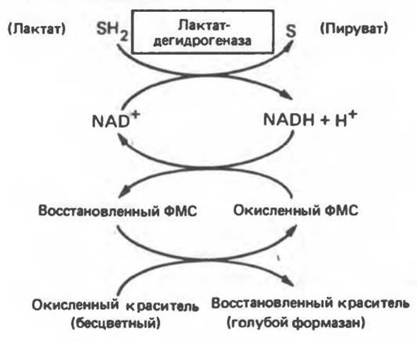
Fig. 7.9. Localization of lactate dehydrogenase on an electrophoregram using a coupled enzyme assay system.
Markert established conditions for the dissociation and reassociation of quaternary structure, thereby clarifying the relationships among lactate dehydrogenase isozymes. Dissociation and reassociation of lactate dehydrogenases I1 and I5 do not lead to The formation of new isozymes, indicating that these two isozymes contain only a single type of protomer. When a mixture of lactate dehydrogenases I1 and I5 was subjected to the same Procedure, forms I2, I3, and I4 also appeared. The ratio of the resulting isozymes corresponds to the subunit composition shown below:

The synthesis of H and M subunits is determined by different genetic loci, and they are differentially expressed in various tissues (e.g., cardiac and Skeletal Muscle).
Last update: 06/08/2026
Editorial and Educational Adaptation: This material has been compiled based on the primary/original source text. The project team performed an editorial review, corrected technical inaccuracies, structured sections, and adapted the content for an educational format.
What was processed:
- elimination of formatting defects (OCR errors, structural breaks, corrupted characters);
- editorial organization of content;
- standardization of terminology in accordance with academic sources;
- verification of factual statements against the original source text.
All mentions of the author, publication year, and origin of the primary text have been preserved in accordance with the source.