Harper's Illustrated Biochemistry, Volume 1 - Murray R. 1993
Structure and Function of Proteins and Enzymes
Proteins: Myoglobin and Hemoglobin
Myoglobin
Biological Function of Myoglobin
Myoglobin is found in red Muscle tissue and is involved in oxygen storage. Under conditions of oxygen deprivation (such as intense physical exertion), oxygen dissociates from its complex with myoglobin and is transported to the Cell/35.html">Mitochondria of muscle Cells, where ATP synthesis takes place via Oxidative Phosphorylation (see Chapter 13).
Primary Structure AND AMINO Acid Distribution
Myoglobin consists of a single polypeptide chain with a Molecular Weight of 17,000; its 153 constituent amino acid residues show no unusual features. However, an analysis of their spatial distribution reveals a distinct pattern: polar residues are located on the molecular surface, while non-polar residues are buried in the interior—a characteristic property of Globular Proteins. Residues containing both polar and non-polar groups (such as Thr, Trp, and Tyr) are oriented with their non-polar groups facing the interior of the globule. With the exception of two Histidine residues involved in oxygen binding, the interior regions of myoglobin contain exclusively non-polar residues (such as Leu, Val, Phe, and Met).
Secondary and Tertiary Structure of Myoglobin
X-ray crystallographic analysis shows that myoglobin is a compact, roughly spherical molecule with dimensions of 4.5 × 3.5 × 2.5 nm (Fig. 6.3). Approximately 75% of the residues form eight right-handed a-helices, each containing from 7 to 20 residues. Starting from the N-terminus, the helices are designated by letters from A to H. The segments connecting the helices are denoted by a combination of two letters indicating the respective helices. Individual residues are assigned a letter denoting the helix in which they reside, followed by a sequence number counted from the N-terminus of that helix. For example, His F8 denotes the eighth residue in helix F, which is a histidine. Residues that are far apart along the primary chain (belonging to different helices, for instance) can be brought into close spatial proximity; notably, the proximal histidine F8 and the distal histidine E7 are located quite close to each other (Fig. 6.3).
A body of evidence indicates that in solution, the secondary and tertiary structures of myoglobin closely resemble those of crystalline myoglobin. In both cases, virtually identical absorption spectra are observed; crystalline myoglobin binds oxygen; and the a-helix content in solution, estimated from optical rotary dispersion and circular dichroism, is consistent with data obtained from X-Ray Diffraction Analysis.
Class="center">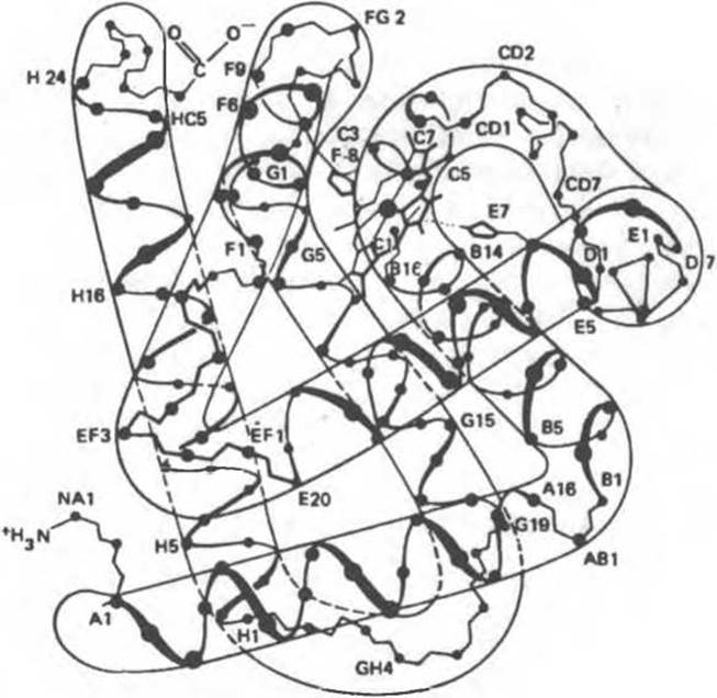
Fig. 6.3. Model of the myoglobin molecule. The contours represent the outlines observed at low resolution. Primarily only the a-carbon atoms and the heme group are shown. (From Dickerson R. E. In: The Proteins, 2nd ed., Vol. 2. Neurath H. (editor). Academic Press, 1964, with kind permission.)
Effect of Heme on Myoglobin Conformation
When the pH is lowered to 3.5, apomyoglobin (heme-free myoglobin) is formed, accompanied by a sharp drop in a-helix content. Subsequent addition of urea to apomyoglobin at neutral pH leads to their nearly complete disappearance. Subsequent removal of urea by dialysis and The addition of heme fully restore the number of a-helices, while the addition of Fe2+ results in the complete recovery of biological (oxygen-binding) activity. Thus, the information encoded in the Introduction/19.html">Primary Structure of apomyoglobin, in the presence of heme, uniquely determines the folding of the protein molecule into its native, biologically active conformation. This important principle applies to other proteins as well: the primary structure of a protein dictates its secondary and tertiary structure.
Spatial Orientation of the Iron Atom, Proximal and Distal Histidine Residues in the Myoglobin Molecule
The heme group in the myoglobin molecule is situated in a crevice between helices E and F; its polar propionate groups are oriented toward The surface of the globule, while the rest of the molecule lies in the interior, surrounded by non-polar residues, with the exception of His F8 and His E7. The fifth coordination position of the iron atom is occupied by the nitrogen atom of the heterocyclic ring of the proximal histidine, His F8 (Fig. 6.4). The distal histidine (His E7) is located on the opposite side of the heme ring, almost directly across from His F8, whereas the sixth coordination position of the iron atom near His E7 remains unoccupied (Fig. 6.4).
Position of the Iron Atom
In deoxymyoglobin, the iron atom is displaced by 0.03 nm out of the plane of the ring toward His F8. In oxymyoglobin, an oxygen atom occupies the sixth coordination position of the iron atom, and the iron atom itself projects only 0.01 nm out of the heme plane. Thus, the oxygenation of myoglobin is accompanied by a Displacement of the iron atom—and consequently of His F8 and its covalently bonded residues—toward the plane of the ring; As a result, this region of the protein globule adopts a new conformation.
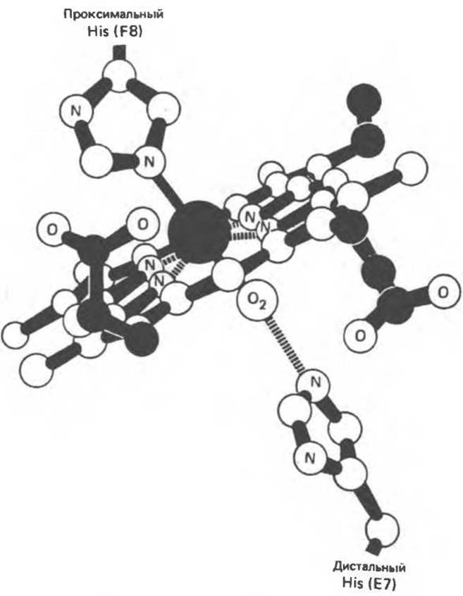
Fig. 6.4. Position of the oxygen molecule in the heme upon oxygenation. The imidazole rings of two crucial histidine residues in the globin chain, positioned adjacent to the iron atom, are also shown. (From Harper H. A. et al., Physiologische Chemie. Springer-Verlag, 1975, with kind permission.)
Ligands
The bond formed between the oxygen atom and the Fe2+ atom during myoglobin oxygenation is oriented perpendicular to the plane of the heme ring. The second oxygen atom is positioned away from the distal histidine, and the bond between the oxygen atoms forms an angle of 121° relative to the heme plane (Fig. 6.5).
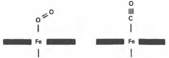
Fig. 6.5. Preferred orientations of oxygen and carbon monoxide molecules bound to the iron atom of isolated heme (dark bars).
Carbon monoxide (CO) binds to isolated heme approximately 25,000 times more tightly than oxygen. Since atmospheric air contains trace amounts of CO, and additional small quantities are produced during normal heme Catabolism, the question arises: Why is the sixth coordination position of iron in myoglobin occupied by an O2 molecule rather than CO? This is due to steric constraints within the myoglobin molecule. When binding to heme, a CO molecule tends to adopt an orientation in which all three atoms (Fe, C, O) lie along a line perpendicular to the plane of the heme ring (Fig. 6.6). While such an orientation is readily achievable for isolated heme, in myoglobin this steric conformation for CO binding is hindered by the distal histidine (Fig. 6.6). Consequently, CO binds in a less favorable configuration, which reduces the affinity of CO for heme by more than two orders of magnitude, making it only about 200 times stronger than the heme–O2 bond. Nevertheless, a small fraction of myoglobin molecules (about 1%) binds CO under normal conditions.
Kinetics of Myoglobin Oxygenation
Why is myoglobin incapable of transporting oxygen, yet highly effective at storing it? The amount of oxygen bound to myoglobin (the "percentage saturation") depends on the oxygen concentration in the immediate microenvironment of the protein molecule (this concentration is expressed as PO2, the partial pressure of oxygen). The relationship between the amount of bound oxygen and PO2 can be represented graphically as the oxygen saturation curve of myoglobin (the oxygen dissociation curve). For myoglobin, the oxygen adsorption isotherm has a hyperbolic shape (Fig. 6.7).
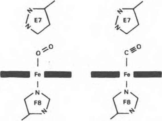
Fig. 6.6. Orientation of oxygen and carbon monoxide molecules bound to the heme iron atom in myoglobin. Distal histidine E7 prevents CO from binding in its preferred orientation, which is at a 90° angle to the plane of the heme ring.
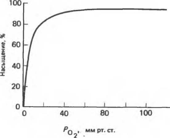
Fig. 6.7. Oxygen saturation curve of myoglobin.
PO2 in the tissue surrounding the pulmonary capillaries is 100 mm Hg, meaning myoglobin could theoretically be saturated with oxygen very efficiently in the Lungs. In venous Blood, PO2 is 40 mm Hg, and in actively working muscle, it is about 20 mm Hg. However, even at a partial pressure of 20 mm Hg, the degree of myoglobin oxygen saturation remains quite high, which is why myoglobin cannot serve as a vehicle for oxygen Transport from the lungs to peripheral Tissues. Nevertheless, during oxygen deprivation associated with intense physical exertion, PO2 in muscle tissue can drop as low as 5 mm Hg; at such low pressures, myoglobin readily releases its bound oxygen, thereby ensuring the oxidative synthesis of ATP in muscle cell mitochondria.
Last update: 06/08/2026
Editorial and Educational Adaptation: This material has been compiled based on the primary/original source text. The project team performed an editorial review, corrected technical inaccuracies, structured sections, and adapted the content for an educational format.
What was processed:
- elimination of formatting defects (OCR errors, structural breaks, corrupted characters);
- editorial organization of content;
- standardization of terminology in accordance with academic sources;
- verification of factual statements against the original source text.
All mentions of the author, publication year, and origin of the primary text have been preserved in accordance with the source.