Fundamentals of Biochemistry - Filippovich, Y. B. 1999
Proteins
Protein Molecule Conformation
The Shape of Protein molecules is determined through the mathematical Processing of data obtained from the ultracentrifugation of protein solutions. Another widely used method for this purpose is Flow Birefringence. It is based on The change in the optical characteristics of a moving protein solution compared to a stationary one: in the former, elongated protein particles align in the direction of fluid flow, which is accompanied by The phenomenon of birefringence. In addition, the shape of protein particles can be established through direct observation using an Electron microscope. Comprehensive data on the shape of protein molecules and their surface topography are provided by X-Ray Diffraction Analysis.
These Methods have revealed that protein particles can be either spherical or highly elongated, taking the form of thread-like structures. In most cases, they have an elongated shape and an asymmetric Structure. The degree of Asymmetry is expressed as The ratio of the particle's long axis, b, to its short axis, a. Data on the degree of asymmetry (b/a) for the molecules of certain Proteins are given in Table 2, and their shapes are illustrated in Fig. 16.
Proteins with an asymmetry degree of 1 (spherical particles) are quite rare. Most commonly, this value ranges from 3 to 6 (ellipsoid or rod-shaped molecules). In some cases, the degree of asymmetry reaches 200 or more (thread-like protein particles). In general, the length of protein molecules with an average molecular weight ranges from several tens to a few nanometers in thickness.
Class="center">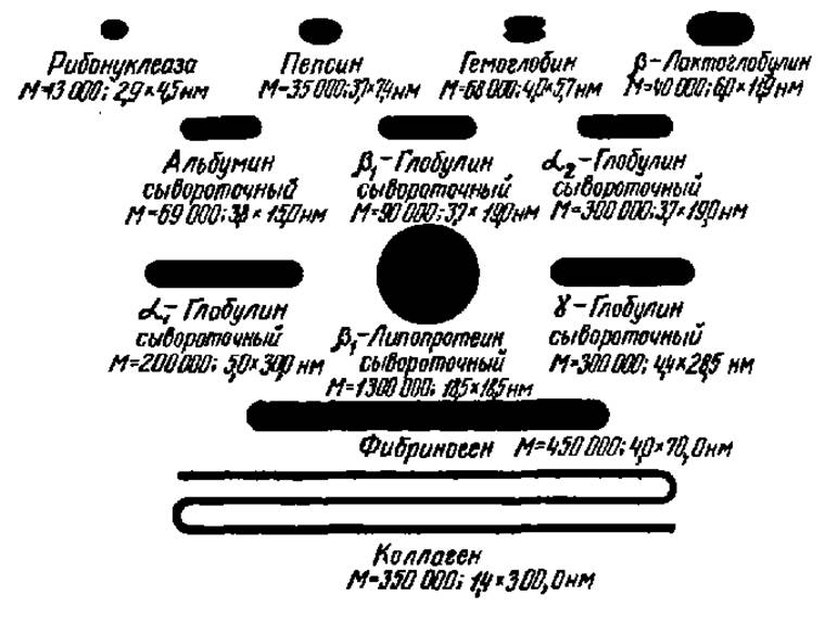
Fig. 16. Shapes of some protein molecules
Below each protein name, its molecular weight in Daltons and Molecular dimensions in nanometers are indicated
Later studies, which detailed the complete structure of certain proteins, showed that protein molecules are asymmetric in all three dimensions. For instance, the molecule of Myoglobin—one of the Muscle tissue proteins (M = 17,600)—has dimensions of 2.5 × 3.5 × 4.5 nm (see pp. 71 and 72). Many proteins, especially Enzymes, exhibit a multilobular structure featuring deep clefts, protrusions, and depressions in their molecules, at the bottom of which functionally active centers are located.
Last update: 06/08/2026
Editorial and Educational Adaptation: This material has been compiled based on the primary/original source text. The project team performed an editorial review, corrected technical inaccuracies, structured sections, and adapted the content for an educational format.
What was processed:
- elimination of formatting defects (OCR errors, structural breaks, corrupted characters);
- editorial organization of content;
- standardization of terminology in accordance with academic sources;
- verification of factual statements against the original source text.
All mentions of the author, publication year, and origin of the primary text have been preserved in accordance with the source.