INVERTEBRATE ZOOLOGY IN THREE VOLUMES — VOLUME 3 — H.I. Shcherbak — 1997
PHYLUM MOLLUSCA (MOLLUSKS)
CLASS SCAPHOPODA (TUSK SHELLS)
This Class comprises a small group of marine benthic Mollusks distributed throughout the World Ocean, particularly in its subtropical and tropical regions from the littoral zone down to depths of 7,000 m. Approximately 300 extant and 700 extinct species are known; a single species, Dentalium novemcostatum, inhabits the Black Sea.
Scaphopods are predominantly small mollusks, ranging from 3 mm to 13 cm in length. The largest of them is the elephant tusk shell (Dentalium elephantium), which reaches up to 15 cm, although the extinct Antalis raymondi is known to have been nearly 60 cm long.
Scaphopods are distinguished by a continuous, narrow, tubular shell open at both ends (Fig. 87), which encloses a large visceral mass; a HEAD bearing numerous tentacles; and a long, narrow FOOT. They possess a Pharynx with a radula and a Liver. The intestine forms an anopedial loop. The Circulatory system is reduced, The Heart lacks atria, and ctenidia are absent. The Central Nervous system is of the scattered-ganglionic type, and Sense Organs are poorly developed. Development proceeds via two larval stages: the trochophore and the veliger.
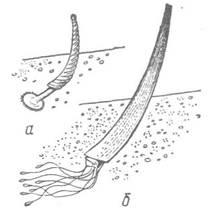
Fig. 87. Class Scaphopoda:
a — Siphonodentalium lofotense; b — Dentalium vulgaris (tusk shell)
The elongated, narrow body of scaphopods consists of a head, a foot, and a trunk, or visceral mass. It is entirely covered by a continuous conical shell with openings at both ends. The head and foot protrude from the wider anterior opening, while only a small appendage of the mantle margin extends from the posterior opening. The shell is slightly curved and resembles an elephant's tusk. It consists of two layers: the prismatic layer (ostracum) and the nacreous layer (hypostracum). The periostracum is undeveloped, which is why the shell is typically white.
The small head appears as an outgrowth bearing the Mouth at its tip, along with two bundles of numerous long, thin tentacles that are thickened at their ends and serve for food collection and as tactile organs. Glandular Cells that secrete mucus are located on the spoon-shaped expansions of the tentacles; this mucus allows small organisms, upon which scaphopods feed, to adhere to the tentacles. The foot is cylindrical, terminating in a cone with a pair of lobes, or it is elongated and worm-like, ending in a jagged disk with a thread-like process in the center (Fig. 87). The foot is well adapted for burrowing in the substrate, hence the class name Scaphopoda (shovel-footed). When moving through the substrate, the foot extends strongly, the lateral lobes fold, and the foot easily penetrates the sediment. The lateral lobes (or disk) then expand and, acting like an anchor, secure the tip of the foot in the sediment. As the foot contracts, the mollusk's body is pulled forward.
The mantle, which covers the visceral mass, fuses around it into a continuous tube. The mantle cavity is long, extending along the ventral side of the body and communicating with the external environment at both ends of the shell. Its expanded anterior part contains the anus and renal openings, whereas the narrow posterior part terminates in an opening at the tip of the shell (Fig. 88). Water is drawn in through the posterior opening and, after supplying oxygen to the Tissues, is expelled through the same opening, carrying away feces, excretory products, and Gametes (during the breeding season). Water Circulation within the mantle cavity is driven by the ciliated epithelium of the mantle.

Fig. 88. Diagram of the anatomy of Dentalium:
1 — tentacles; 2 — mouth; 3 — cerebral ganglion; 4 — Stomach; 5 — Pericardium with heart; 6 — Kidney; 7 — gonad; 8 — upper mantle opening; 9 — liver; 10 — anus; 11 — shell; 12 — mantle; 13 — statocyst; 14 — pedal ganglion; 15 — foot
The Digestive System of scaphopods is represented by all the subdivisions characteristic of mollusks. They possess a pharynx with a horseshoe-shaped jaw, a radula, an Esophagus, a stomach, a bilobed liver, and Midgut and Hindgut sections. The intestine forms a loop (the anopedial loop); the anus is situated near the anterior end of the body. Scaphopods feed on small benthic invertebrates—foraminiferans, ostracods, juvenile bivalves, etc.—gathering them from the substrate using their adhesive tentacles.
The excretory organs are a pair of Kidneys that do not communicate with the pericardium but open solely into the mantle cavity. The excretory ducts of the kidneys also serve to discharge reproductive products (see below).
The circulatory system is reduced. Blood Vessels are absent; however, There is a heart enclosed within a pericardium, consisting of a single ventricle. Blood flows exclusively through lacunae and sinuses.
Ctenidia are absent, and Respiration is carried out via thin mantle folds located on the walls of the mantle cavity.
The central nervous system consists of four pairs of ganglia connected by connectives. The paired cerebral and pleural ganglia lie closely appressed to one another, while the pedal and visceral ganglia are located in the foot and near the anus, respectively. Buccal ganglia innervating the pharynx are also present.
Sense organs are poorly developed. Eyes are absent; there is a single pair of equilibrium organs, the statocysts. The oral tentacles function as tactile organs, while the subradular organ located in the pharynx serves as a chemoreceptor.
Scaphopods are dioecious; the gonad is unpaired and lacks dedicated genital ducts, opening instead into the right kidney, from which mature gametes are discharged via its excretory duct. Fertilization is external.
Embryonic development follows the pattern typical of mollusks; a trochophore larva hatches from the egg. Subsequently, a shallow depression—the shell gland—develops on the DORSAL SIDE OF the larva. The upper hemisphere of the trochophore flattens, transforming into a disk-shaped velum, while the posterior region begins to elongate. Two longitudinal Skin folds—the mantle rudiments—appear on the sides of the body, and their margins fuse on the ventral side (Fig. 89). Following the mantle, the shell also acquires a tubular shape, being secreted by the mantle and fused on the ventral side. The rudiment of the foot forms posterior to the mouth; this larval stage is termed the veliger. After a few days of planktonic life, metamorphosis begins: the velum and apical organ regress, and the larva settles to the bottom.

Fig. 89. Development of Dentalium:
a, b — early and late veligers; 1 — velum; 2 — mantle; 3 — shell; 4 — foot rudiment
Scaphopods primarily inhabit shallow waters, living buried in soft sediments with only the posterior end of the shell protruding above the surface.
Last update: 13/08/2026
Editorial and Educational Adaptation: This material has been compiled based on the primary/original source text. The project team performed an editorial review, corrected technical inaccuracies, structured sections, and adapted the content for an educational format.
What was processed:
- elimination of formatting defects (OCR errors, structural breaks, corrupted characters);
- editorial organization of content;
- standardization of terminology in accordance with academic sources;
- verification of factual statements against the original source text.
All mentions of the author, publication year, and origin of the primary text have been preserved in accordance with the source.