INVERTEBRATE ZOOLOGY IN THREE BOOKS - BOOK 2 - H.I. Shcherbak - 1996
PHYLUM ECHIURIDA
CLASS ECHIURIDA
サイズの小さいエхиурид (with an extended proboscis) range from 3 to 200 cm, while trunk dimensions vary from a few millimeters to 20–30 cm.The trunk in most echiurans (Fig. 42) is elongated and cylindrical, although some species have a shorter, sac-like or even spherical trunk. It bears several groups of chitinous setae: two robust setae are located on the ventral side just behind the Mouth (known as genital setae), and one or two rings of tiny setae are found at the posterior end of the body (some setae may occasionally be absent). Extending forward from the trunk is a body region known as the proboscis. Its length varies within a wide range: in some species, it does not exceed the length of the trunk, while in others, it may be tens of times longer. The proboscis cannot be retracted into the trunk, but it is capable of strong contraction and elongation. The anterior end of the proboscis sometimes expands into a large lobe, which may be continuous in some species or divided into two parts in others. Its ventral surface is slightly concave and ciliated. Frequently, the proboscis is rolled longitudinally into a funnel, at the bottom of which the mouth is situated (Echiurus echiurus).
Class="center">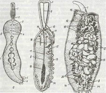
Fig. 42. Echiurus echiurus:
a – general appearance; b – diagram of internal Structure; c – dorsal dissection; 1 – proboscis; 2 – mouth;
3 – genital setae; 4 – genital pores; 5 – anal setae; 6 – anus; 7 – intestinal loops visible through the body wall; 8 – ventral nerve cord; 9 – dorsal Blood vessel; 10 – Pharynx; 11 – Esophagus; 12 – siphon; 13 – midgut loops; 14 – anal sac; 15 – anus; 16 – gonad; 17 – ventral blood vessel; 18 – nephridiostomes; 19 – sacs of ventral setae;
20 – mesenteries
The body of echiurans is covered by a cuticle, beneath which lies a single-layered epithelium. Cilia persist only on the proboscis. In deep-sea forms, the cuticle is thin and transparent, whereas in shallow-Water species, it is denser and covered with thickenings called papillae, which are believed to serve a sensory function. Echiurans are often colored yellow, various shades of red, or green.
Beneath the epithelium lies a muscular body wall composed of circular, longitudinal, and oblique Muscle layers. Internally, it is lined by peritoneal epithelium, which encloses the coelom. The same epithelium surrounds the intestine, forming numerous mesenterial strands and folds that connect with the peritoneal epithelium of the body wall and support the intestinal loops (Fig. 42, c).
The body cavity is an unsegmented coelom that occupies the entire trunk. Its anterior wall (Diaphragm) is located in the anterior part of the trunk, anterior to the genital setae. The diaphragm has two apertures: the intestine passes through one, while the other, a narrow slit, connects the coelom with the cavity of the proboscis. The latter continues into three very narrow lacunae extending the entire length of the proboscis. The exact Nature of the proboscis cavity remains debated; some scientists consider it an extension of the coelom, while others view it as a primary body cavity (schizocoel).
Echiurans are sessile animals that inhabit the substrate with only their proboscis protruding. When the trunk Muscles contract, coelomic fluid is forced under pressure into the channels of the proboscis, causing it to elongate and become rigid; upon muscle relaxation, internal pressure decreases, and the proboscis deflates.
The Digestive System (Fig. 42, b, c) is adapted for feeding on small detritus particles, microorganisms, and the like. The mouth is situated on the ventral side at the boundary between the trunk and the proboscis. Cilia covering the groove on the ventral side of the proboscis sweep food toward the oral aperture. The intestine is extremely long and coiled, measuring up to 10 times the body length. It comprises foregut, midgut, and hindgut regions. The ectodermal foregut includes the Oral Cavity, pharynx, esophagus, and crop. The entodermal midgut features a so-called siphon—a slender tube branching off from the intestine and running alongside this region to its posterior border. The ectodermal hindgut is short and bears a dilation into which a pair of large anal sacs empty.
The excretory system is represented by several Organs. Larvae possess a pair of protonephridia, which degenerate during metamorphosis. In adults, two elongated anal sacs open into the hindgut. Each sac is covered with numerous (from 10 to 300) funnels. By means of their expanded, ciliated ends, each funnel opens into the body cavity, while the narrowed end opens into the sac. Some researchers suggest that these funnels are modified metanephridia. In addition to excretion, the anal sacs perform a respiratory function. Furthermore, in the anterior region of the trunk, there are one or more (up to 200 pairs) nephridiostomes (nephromixia), which primarily serve to discharge Gametes rather than metabolic wastes. All nephromixia open externally via a common pore (nephridiopore) located on the ventral side posterior to the mouth. Occasionally, the number of pores increases to four.
The Circulatory system is closed. It consists of a long ventral vessel running along the entire body above the ventral nerve cord. Posterior to the mouth, it bifurcates, with both branches passing inside the proboscis; in the anterior part of the proboscis, they reunite into a single dorsal vessel that emerges from the proboscis, runs above the esophagus, encircles it as a vascular ring, and connects with the ventral vessel. The vascular ring around the esophagus has muscular walls and pulsates, thus functioning as a Heart. The blood is colorless and contains amoeboid Cells.
Echiurans respire through their entire body surface.
The Nervous system consists of a strongly elongated nerve ring that extends along the entire proboscis to its anterior tip, and a ventral nerve cord. The latter connects with the nerve ring behind the mouth and lies within the body cavity, being attached to the body wall by a narrow mesentery. Ganglia are completely absent.
Sense Organs are poorly developed. Besides Skin papillae, the tip of the proboscis bears chemoreceptors and specialized receptors responsive to water currents. Irritation of the latter causes the proboscis to retract.
Echiurans are dioecious. Males and females generally share the same Morphology, and pronounced Sexual Dimorphism is observed in only a few species. For instance, in members of the family Bonellidae, males are dwarf (1–3 mm) and lack a mouth, anus, and circulatory system; their bodies are covered with ciliated epithelium. These males inhabit the nephromixia of the females, which is why they were long mistaken for parasites.
Gonads in echiurans appear as groups of cells located within the peritoneal epithelium along the posterior part of the ventral blood vessel. Germ Cells are released into the coelom, where they mature while floating in the coelomic fluid; they are expelled to the exterior via the nephromixia.
Fertilization is typically external, with eggs fertilized by sperm upon release; during this process, the worms pair up via their nephridiopores using genital setae.
Cleavage is spiral and determinate. Embryonic development leads to The formation of a trochophore larva (Fig. 43). It features an apical plate at the animal pole and several rings of cilia, the prototroch, and in some species, a metatroch and telotroch. The mouth is located near the equator, ventral to the prototroch. The trochophore possesses a primary body cavity, a pair of mesodermal bands representing the rudiments of the mesoderm, and a pair of protonephridia.
The larva swims in the water Column. Metamorphosis occurs gradually: the upper hemisphere of the trochophore progressively assumes the shape of the preoral lobe (proboscis), while the lower hemisphere grows extensively to become the trunk of the adult animal. No specialized growth zone is formed during this process, and the body enlarges more or less uniformly in all its parts. The mesodermal bands do not become segmented; instead, they expand, fuse with one another, and subsequently split into two layers. The inner layer envelops the gut, while the outer layer lines the body wall, enclosing a continuous coelomic cavity between them. Following metamorphosis, the larva settles to the bottom.

Fig. 43. Trochophore of Echiurus echiurus, ventral view:
1 - apical plate; 2 - circumoesophageal connective; 3 - prototroch; 4 - metatroch; 5 - mouth; 6 - protonephridium; 7 - anus;
8 - anal sac rudiment; 9 - external integumentary folding; 10 - ventral nerve cord; 11 — gut
A very fascinating metamorphosis is observed in echiurans of the family Bonellidae, which, as mentioned previously, feature dwarf males (Fig. 44). Special studies have shown that the Transformation of a bonellid larva into a male or a female depends entirely on environmental conditions. When larvae are kept in an aquarium without adult females, they develop into females very slowly, over the course of several weeks. These females grow sluggishly, reaching sexual maturity and a length of 10–12 cm only after a year. If an adult female is introduced into the aquarium with the larvae, metamorphosis proceeds differently depending on where the larva settles: if it attaches to the female's proboscis, its metamorphosis takes place within a day, transforming it into a male that becomes sexually mature in 4–5 days. Residing within the nephromixia of the females, these males fertilize the eggs passing through them. Larvae also transform into males if only the proboscis of a female, or even an extract of the proboscises, is placed in the aquarium. It is believed that the female proboscis contains a hormone-like substance with a specific effect on the larvae. Thus, The Development of bonellids represents one of the very rare instances among animals where sex is determined after egg fertilization. However, in 10% of cases, larval development deviates from this pattern, suggesting that genetic factors also play a role in Sex Determination.
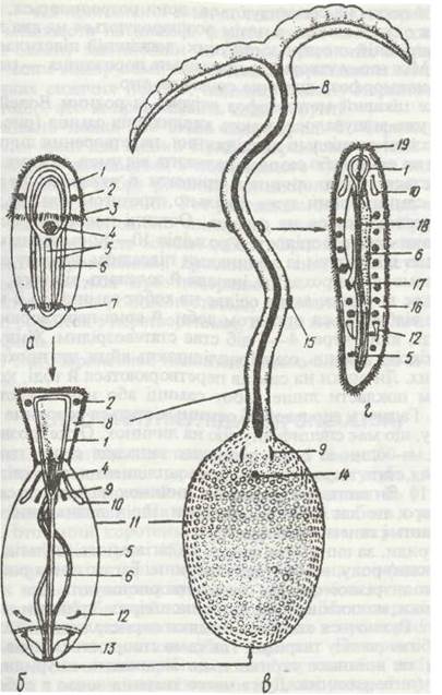
Fig. 44. Metamorphosis and phenotypic Sex determination in Bonellia viridis:
a - free-swimming larva; b - older female larva; c - adult female; d - male; 1 - circumoesophageal nerve ring; 2 - eyes; 3 - prototroch; 4 - foregut; 5 - ventral nerve cord; 6 - midgut; 7 - anal ciliated wreath; 8 - proboscis; 9 - setae; 10 - protonephridium; 11 - trunk; 12 - metanephridium; 13 - anal sac; 14 - female genital opening; 15 - larva on the female's proboscis; 16 - coelom with developing sperm cells; 17 - male genital funnel; 18 - sperm duct; 19 - male genital opening
With the exception of a single pelagic genus, echiurans are benthic animals. Many of them burrow various types of tunnels in the sediment, while others inhabit mollusk shells, echinoderm skeletons, rock crevices, and similar shelters. Echiurans move by means of peristaltic waves rippling through their bodies. This same mechanism generates water currents in their burrows and retreats, ensuring a continuous supply of fresh water for Respiration. Very frequently, the animal conceals its trunk within the substrate while using its long, mobile proboscis to forage for food in the surrounding area. The proboscis often falls prey to various predators, but it possesses a remarkable capacity for regeneration.
Last update: 13/08/2026
Editorial and Educational Adaptation: This material has been compiled based on the primary/original source text. The project team performed an editorial review, corrected technical inaccuracies, structured sections, and adapted the content for an educational format.
What was processed:
- elimination of formatting defects (OCR errors, structural breaks, corrupted characters);
- editorial organization of content;
- standardization of terminology in accordance with academic sources;
- verification of factual statements against the original source text.
All mentions of the author, publication year, and origin of the primary text have been preserved in accordance with the source.