IMMUNOLOGY - Roit I. - Mir 2000
Chapter 14. Immunological Tolerance
CENTRAL THYMIC TOLERANCE TO AUTOANTIGENS
The Thymus is the site where T Cells differentiate from precursor cells that carry germline T-Cell receptor (TCR) genes. During lymphoid development within the thymus, these genes undergo rearrangement, enabling T cells to express TCRs capable of recognizing degradation products of Antigens, or Peptides bound in the peptide-binding groove of MHC molecules—which are encoded by the GENES OF THE Major Histocompatibility Complex (MHC) (see Chapters 7 and 9).
The thymus selects for T cells bearing receptors that can bind antigens complexed with MHC molecules, while deleting cells with high avidity for self-antigens.
The high proliferation rate of thymocytes is matched by their massive cell death: the overwhelming majority of so-called double-positive (CD4+CD8+) thymocytes perish within the thymus. The reasons for this include aberrant TCR Gene rearrangement (the generation of 'useless' receptors), negative Selection, and failure to pass positive selection. A prerequisite for positive selection is a moderate affinity of T cells for polymorphic regions of MHC molecules associated with peptides; such cells survive (Fig. 14.2). They interact with MHC molecules On the surface of cortical epithelial cells, an engagement that is thought to rescue T cells from programmed cell death (apoptosis). Positive selection ensures the generation of mature T cells capable of recognizing only those peptides that are cradled within the binding groove of self-MHC molecules. This phenomenon of restricted recognition is known as MHC restriction. However, positive selection does not prevent the maturation of T cells bearing receptors with high affinity for both self-peptides and MHC molecules. Therefore, mechanisms of negative selection must exist to silence such highly autoreactive cells.
Class="center">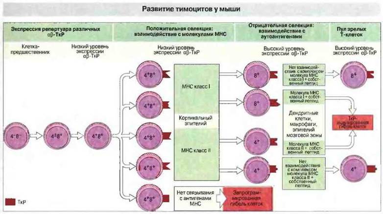
Fig. 14.2. Thymocyte precursors in the thymic cortex differentiate into 'double-positive' cells expressing low levels of αβ-TCRs. These cells undergo positive selection by interacting with self-MHC class I or II molecules on cortical epithelium. Unselected cells (the majority) die via programmed self-destruction (apoptosis). Positively selected cells downregulate one of their coreceptor molecules (CD4 or CD8). Finally, autoreactive cells are eliminated through their interaction with self-peptides presented on cells at the corticomedullary junction and in the thymic medulla.
Mechanisms of positive and negative selection
Since both positive and negative selection involve the recognition of self-peptides associated with self-MHC molecules, the question arises as to how signals transduced through the exact same TCR can drive two such divergent outcomes. Two main theories have been proposed to explain this.
✵ The quantitative model posits that low numbers of peptide-MHC complexes induce positive selection, whereas high numbers trigger negative selection.
✵ The core premise of the 'qualitative' model is that, upon binding to the same T-cell receptor, different peptide-MHC complexes deliver qualitatively distinct signals that result in either positive or negative selection.
Support for the quantitative model comes from experiments using Transgenic Mice expressing a TCR specific for myelin basic protein, an autoantigen of the myelin sheath surrounding axons. In one mouse strain, the natural peptide epitope recognized by these T-specific cells binds its restricting MHC class II molecule with very low affinity. Consequently, epitope-specific T cells escape negative selection in the thymus. However, if these transgenic mice carrying the same TCR are administered a high-affinity analog of this peptide, immature thymocytes undergo negative selection via apoptotic cell death (Fig. 14.3). Evidently, cell fate—whether death or survival—is dictated by the number of peptide-MHC complexes presented to developing T cells. Self-epitopes that form more stable complexes with MHC Proteins are far more likely to induce negative selection.
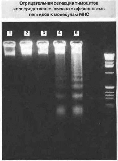
Fig. 14.3. This DNA-stained agarose gel demonstrates varying degrees of apoptosis (DNA fragmentation) in thymocytes from transgenic mice (expressing TCRs specific for the Ac1-9 peptides of myelin basic protein) in the presence of peptides showing increasing affinity for MHC class II molecules.
1. Irrelevant peptide.
2. Native low-affinity peptide (Ac1-9).
3. Intermediate-affinity MHC analog (Ac1-9 [4A]).
4. High-affinity MHC analog (Ac1-9 [4V]).
5. Another high-affinity MHC analog (Ac1-9 [4Y]).
The qualitative theory was put forward to explain findings from fetal thymic organ cultures. These experiments demonstrated that peptide epitopes modified at amino acid residues responsible for contacting the T-cell receptor—known as TCR antagonists—promote more robust positive selection than the wild-type epitope (agonist). Nevertheless, these two theories are not mutually exclusive. Modern experimental techniques now allow the measurement of binding affinities between purified TCR molecules and purified MHC molecules loaded with peptide agonists or antagonists. Using these Methods, it has been established that TCRs form unstable interactions with peptide-antagonist-MHC complexes that induce positive selection in thymic organ cultures (Fig. 14.4). Conversely, the exact same TCR binds relatively stably to the agonist-MHC complex, which under identical experimental conditions leads to negative selection. These findings bridge the quantitative and qualitative models, aligning with a kinetic model in which the induction of positive versus negative selection is determined by the off-rate (dissociation rate) of the TCR from the peptide-MHC complex. The overall avidity of T-cell interactions with antigen-presenting cells (APCs) in the thymus (Fig. 14.5) is governed by the following factors:
✵ the number of peptide-MHC complexes, which depends on the affinity of the epitope for the relevant MHC molecule;
✵ the density of TCRs and corresponding coreceptor molecules expressed on the T-cell surface; and
✵ the affinity of the specific TCR for its Ligand.
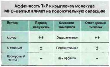
Fig. 14.4. The affinity of soluble TCR for complexes of various peptides and corresponding restriction elements (MHC antigens) can be determined using biophysical methods, such as surface plasmon Resonance. There is a direct correlation between the half-life of the TCR–MHC-peptide complex bond and the response of mature T cells expressing the same receptor (i.e., agonist > antagonist > irrelevant peptide). However, in fetal organ thymic cultures, The addition of an agonist peptide causes the deletion of developing cells (negative selection), whereas an antagonist peptide promotes positive selection. This demonstrates that low-avidity interactions stimulate positive selection, while high-avidity interactions induce negative selection.
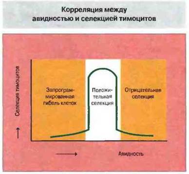
Fig. 14.5. The avidity of T-cell interaction with an antigenic peptide presented on an APC depends on the expression level (concentration) of the MHC molecule–peptide complex [MHC molecule + peptide] on the APC, as well as on the affinity and surface expression (concentration) of TCRs and coreceptor molecules [TCR + coreceptor molecules] on T cells. [MHC molecule + peptide] depends on the affinity of the peptide for the MHC molecule and the Stability of the resulting complex. [TCR + coreceptor molecules] is determined by the affinity of the individual TCR for the peptide–MHC molecule complex, the level of TCR expression on The Cell surface, and the corresponding level of coreceptor molecule expression.
Positive selection and T-cell commitment occur at a specific stage of their development
Why do thymocytes lose one of their coreceptor molecules during development and become either CD4+ or CD8+ cells? Positive selection affects only those immature thymocytes that bear TCRs of appropriate Specificity and still express both coreceptors. Positively selected thymocytes differentiate into so-called single-positive cells, either CD4+ or CD8+. Upon commitment to a particular cell Lineage, the Synthesis of the alternative coreceptor molecule is selectively turned off. In mice, this selective shutdown occurs when cells bearing both coreceptors receive the signals necessary for positive selection (Fig. 14.2).
The timing and Location of negative selection depend on various factors
These factors include the availability of self-antigens to developing T cells, the combined avidity of TCRs and accessory molecules (CD8 or CD4) for the self-antigen MHC–self-peptide complex, and The Nature of the cells responsible for deletion. Negative selection does not require specialized APCs; under normal conditions, this function is performed by thymic dendritic cells or macrophages, which are localized predominantly at the corticomedullary junction. They carry a high density of class I and II MHC molecules on their surface, thereby ensuring the capture of T cells with high avidity for self-peptides (Fig. 14.6). Certain cortical and medullary epithelial cells may also participate in negative selection. Thymocytes themselves are also capable of mediating deletion (Fig. 14.6).
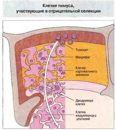
Fig. 14.6. The deleting population comprises Bone Marrow-derived macrophages or dendritic cells located predominantly at the corticomedullary junction. Other cells may also participate in deletion, notably thymocytes themselves acting via "veto" activity, as well as certain types of thymic epithelial cells, likely localized in the medulla.
Negative selection involves a specialized death signal
There are several mechanisms of apoptotic death in mature lymphocytes. Signals inducing apoptosis can be transmitted via Fas, CTLA-4, or TNF receptors (see below). The process of negative selection in double-positive thymocytes (CD4+CD8+) is unique in that it does not depend on any of these pathways.
Last update: 13/08/2026
Editorial and Educational Adaptation: This material has been compiled based on the primary/original source text. The project team performed an editorial review, corrected technical inaccuracies, structured sections, and adapted the content for an educational format.
What was processed:
- elimination of formatting defects (OCR errors, structural breaks, corrupted characters);
- editorial organization of content;
- standardization of terminology in accordance with academic sources;
- verification of factual statements against the original source text.
All mentions of the author, publication year, and origin of the primary text have been preserved in accordance with the source.