IMMUNOLOGY - Roitt I. - Mosby 2000
Chapter 12. Ontogeny of the Immune System
■ The majority of immune Cells originate from hematopoietic stem cells.
■ The Development of distinct Cell lineages (differentiation pathways) depends on intercellular interactions and cytokines.
■ Lymphoid stem cells develop and mature within Primary Lymphoid Organs, a process known as lymphopoiesis.
■ T lymphocytes undergo positive and negative Selection during their development in the Thymus.
■ Mammalian B cells develop primarily in the fetal Liver and subsequently in the Bone Marrow after birth, a process that continues throughout the Organism's lifespan. B cells also undergo selection at their sites of development.
■ The antibody repertoire present in adults is established during lymphopoiesis through the recombination of Gene segments encoding TCR and Ig.
■ Germinal centers are sites of oligoclonal B-cell proliferation, antibody isotype switching, affinity maturation, and immunological memory formation.
The effective functioning of The Immune System relies on the interplay of numerous cellular and humoral components that mature at varying rates during the pre- and postnatal periods. Many cells involved in the Immune Response derive from undifferentiated hematopoietic stem cells (HSCs). Influenced by microenvironmental factors—such as cell-to-cell contacts and the presence of soluble or membrane-bound cytokines—HSCs differentiate along multiple pathways (Fig. 12.1).
Class="center">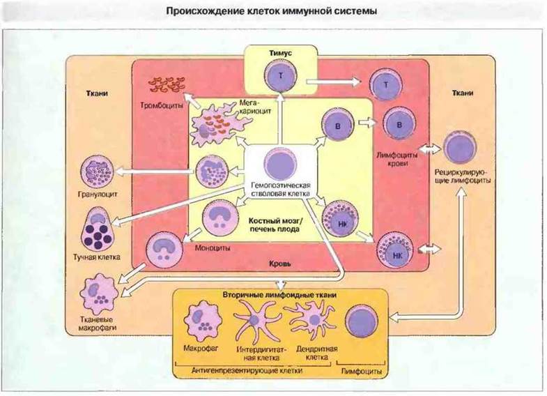
Fig. 12.1. All cells illustrated in the diagram originate from a hematopoietic stem cell. Platelets, produced by megakaryocytes, enter the bloodstream. Granulocytes and monocytes migrate from the Circulation into Tissues. Mast cells are present in all tissues. In mammals, B cells mature in the fetal liver and bone marrow, whereas T cells mature in the thymus. Large granular lymphocytes with natural killer (NK) cell activity are likely generated in the bone marrow. Lymphocytes migrate from the bloodstream, pass through secondary lymphoid tissues, and recirculate back into the Blood. Interdigitating and dendritic cells act as antigen-presenting cells within secondary lymphoid tissues.
In mammals, during embryonic development, HSCs are located in the yolk sac, liver, Spleen, and bone marrow. After birth and throughout adult life, they typically persist only in the bone marrow. These self-renewing HSCs, driven by diverse local growth and differentiation factors within hematopoietic niches, give rise to most, if not all, Cells of the immune system.
HSCs give rise to four main lineages of differentiation:
✵ erythroid (erythrocytes),
✵ megakaryocytic (platelets),
✵ myeloid (granulocytes and mononuclear phagocytes), and
✵ lymphoid (lymphocytes). Antigen-presenting cells develop predominantly, though not exclusively, from myeloid progenitor cells. Myeloid and lymphoid Lineage cells are the most critical for immune system function.
MYELOID CELLS
In humans, myelopoiesis begins in the liver at approximately the 6th week of gestation. In vitro studies of colony growth from individual stem cells have demonstrated that the earliest HSC-derived progenitor is a colony-forming unit (CFU) capable of generating granulocytes, erythrocytes, monocytes, and megakaryocytes (CFU-GEMM). The maturation of these cells is driven by colony-stimulating factors (CSFs) and several interleukins, including IL-1, IL-3, IL-4, IL-5, and IL-6 (Fig. 12.2). All of these factors play a vital role in the upregulation (stimulation) of hematopoiesis and are produced primarily by bone marrow stromal cells, as well as by mature forms of differentiated myeloid and lymphoid cells. Other cytokines (such as TGF-β) can mediate the downregulation (suppression) of hematopoiesis.
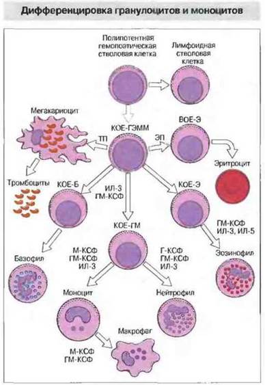
Fig. 12.2. Pluripotent hematopoietic stem cells give rise to colony-forming units (CFUs). CFU-GEMM can generate all Blood Cells except lymphocytes. The derivation of any of the five cell types from the stem cell [megakaryocytes; erythrocytes—via a progenitor stage termed the burst-forming unit-erythroid (BFU-E); basophils, neutrophils, or eosinophils] is induced by IL-3 and granulocyte-macrophage colony-stimulating factor (GM-CSF); these same factors are required for the further differentiation of granulocytes and monocytes. The production of eosinophils (E) and CFU-E is stimulated by IL-5. Neutrophils and monocytes develop from CFU-GM under METABOLISM/18.html">The Influence of G-CSF and M-CSF, respectively. Both of these factors, alongside other cytokines (including IL-1, IL-4, and IL-6), stimulate the differentiation of monocytes into macrophages. Megakaryocyte production is stimulated by thrombopoietin (TPO). (B = basophil; EPO = Erythropoietin; G = granulocyte; M = monocyte.)
Neutrophils and monocytes develop from common progenitor cells
Neutrophil development. The common progenitor for neutrophils (a type of granulocyte) and mononuclear phagocytes (macrophages) is the CFU-GM. As they differentiate into neutrophils, the cells progress through several morphological stages. Myeloblasts give rise to promyelocytes and subsequently myelocytes, which mature and enter the circulation as neutrophils. The unilinear differentiation of CFU-GM cells into mature neutrophils is governed by the surface expression of receptors for specific growth and differentiation factors at various Stages of development.
As granulocytes mature, specific surface differentiation markers appear or disappear on their cell membranes (Fig. 12.3). For instance, CFU-GM cells express MHC class II molecules and the CD38 marker, both of which are absent on mature neutrophils. Other surface molecules expressed during differentiation include CD13 and CD14.
(present in low concentration), CD15 (Lewis blood group X-determinant), CD29 (β1-integrin), VLA-4 (CD49d, α-chain), leukocyte Integrins CD11a, b, c, and αD in association with β2-chains CD18, Complement receptors, and Fcγ-receptors (CD16) (see Fig. 2.42).
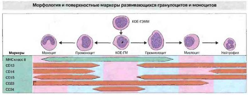
Fig. 12.3. Cells of the monocytic and neutrophilic differentiation lineages originate from a common progenitor cell, CFU-GM. Differentiation along each of these pathways is accompanied by the loss of the CD34 marker. CD33 is retained on monocytes, whereas mature neutrophils lack these molecules, as well as MHC class II molecules. The CD14 marker is expressed on monocytes, but only weakly on certain granulocytes, possibly upon activation.
Assessing the functional activity of granulocytes at various stages of maturation is challenging, but it appears that only mature cells possess full functional capacity. A body of evidence indicates that neutrophil activity, as determined by phagocytosis or chemotaxis, is lower in the fetus than in the mature organism. However, this may be partly due to a lower opsonin content in fetal serum rather than intrinsic Properties of the cells themselves. To acquire activity, neutrophils require direct interaction with microorganisms, or with cytokines produced during the Immune Response to an antigen (or both together) in the presence of opsonins. This may limit neutrophil activity in the Cytology/cytology/16.html">Early stages of development. Activation of neutrophils by cytokines and chemokines is also a prerequisite for their migration from the blood into tissues.
Monocyte formation. During monocytic differentiation from CFU-GM, proliferating monoblasts are initially formed. These differentiate into promonocytes and, finally, into mature blood monocytes. Circulating monocytes are thought to serve as a renewable pool for the generation of tissue macrophages, such as lung macrophages. Various Forms of macrophages constitute the mononuclear phagocyte system (see Chapter 2).
Mature neutrophils and monocytes/macrophages lack CD34 and other early differentiation markers. However, unlike neutrophils, monocytes continue to express high levels of MHC class II molecules (Fig. 12.3), which are essential for antigen presentation to T cells. Monocytes also synthesize many of the surface molecules characteristic of mature neutrophils (see Fig. 2.28).
Determining the functional capabilities of monocytes across differentiation stages is, as with granulocytes, quite difficult. Nevertheless, in vitro studies of certain myeloid tumors, whose cells presumably represent monocytes at various stages of differentiation, indicate that both phagocytic activity and Fc receptor-mediated cytotoxicity reach optimal levels only at the mature macrophage stage. In both newborns and adults, monocytes produce the cytokine IL-1 with equal efficiency, but in newborns, this function is less responsive to upregulation by IFN-γ than in adults.
Dendritic cells develop from bone marrow stem cells
Most classical antigen-presenting cells (APCs), including macrophages, Langerhans cells, interdigitating cells, and dendritic cells, are already present in the body at birth. Most likely, the bulk of them originate from bone marrow stem cells. They may derive from the same CD34+ stem cell (CFU-GEMM). The morphological, cytochemical, and Functional Characteristics of different APCs must then be determined by subsequent microenvironmental influences, such as cytokines. Another possibility is that APCs originate from different stem cells along separate differentiation pathways. An important exception is follicular dendritic cells (FDCs), which are localized in germinal centers within secondary lymphoid follicles and presumably originate from mesenchymal cells. FDCs are present in the primary follicles of peripheral lymphoid tissues at birth. Unlike other APCs, they are non-motile.
APCs are present in the thymus at very early stages of development, and their involvement in MHC restriction and T-cell selection indicates that at least some of them achieve full maturity by this time. However, APC activity in early developmental stages is clearly suboptimal. In newborn rats, for example, antibody production against sheep red blood cells occurs only with the simultaneous administration of APCs from adult rats (Fig. 12.4).
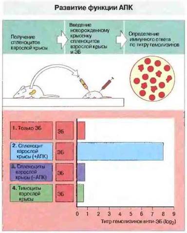
Fig. 12.4. Development of antigen-presenting cell function in ANTIGEN Processing AND presentation. In this experiment, newborn rats were administered: 1) sheep red blood cells (SRBCs) only, 2) SRBCs + spleen cells (containing APCs) from adult rats, 3) SRBCs + APC-depleted spleen cells, or 4) SRBCs + mature thymocytes. In all cases, the adult rats belonged to the same strain as the newborns. The HUMORAL IMMUNE RESPONSE—the appearance of Antibodies—was recorded in the rats of each group. Newborn rats injected with SRBCs alone failed to produce antibodies against SRBC Antigens. However, upon simultaneous administration of adult rat splenocytes (option 2), an immune response developed. Neither mature splenocytes lacking APCs nor thymocytes alone induced antibody production. Consequently, APCs from newborns are incapable of efficiently processing and presenting SRBC antigens.
Last update: 13/08/2026
Editorial and Educational Adaptation: This material has been compiled based on the primary/original source text. The project team performed an editorial review, corrected technical inaccuracies, structured sections, and adapted the content for an educational format.
What was processed:
- elimination of formatting defects (OCR errors, structural breaks, corrupted characters);
- editorial organization of content;
- standardization of terminology in accordance with academic sources;
- verification of factual statements against the original source text.
All mentions of the author, publication year, and origin of the primary text have been preserved in accordance with the source.