Introduction to Molecular Biology: From Cells to Atoms - Anthony Rees, Michael Sternberg 2002
Carbohydrates and Lipids
Structure of Polysaccharides
Class="center">Introduction/introduction.files/image080.jpg" width="621"/>
Fig. 31.1.
Polysaccharides are long chains of Monosaccharides linked together by glycosidic bonds. They often possess a linear Structure, but can also be branched. In both plants and animals, polysaccharides play structural roles and serve as energy reserves. The most common monosaccharide found in polysaccharides is the six-carbon sugar (hexose) D-glucose. Due to the various ways hexoses can link (types of glycosidic bonds) and their diverse functional groups, The structure of polysaccharides is highly varied. We will examine only five of the many existing polysaccharides.
D-Glucose is a six-carbon monosaccharide; its structure is shown in Fig. 31.2. The —OH group at carbon atom 1 is directed either upward (i.e., in the same direction as carbon atom 6) to form ß-D-glucose, or downward to form a-D-glucose. The difference between these related sugars plays a crucial role when they form polysaccharides. Hexoses (such as D-glucose) can exist in two stable Conformations representing two Variants of the chair conformation (Chap. 14): C1 and 1C. In the C1 state, the bulky OH groups are parallel to the plane of the sugar ring and point outward; such groups are said to be in an equatorial orientation. In the 1C state, these groups point upward and downward relative to the ring, meaning they have an axial orientation. Since oxygen is a relatively large atom, the C1 conformation is more favorable because it keeps large atoms well separated from one another.
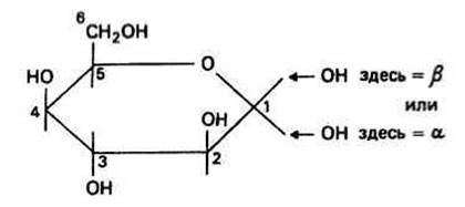
Fig. 31.2.
A glycosidic bond is formed through the interaction of two monosaccharides. It is an ether bond whose formation is accompanied by the release of a Water molecule (Fig. 31.1). There are various ways in which glycosidic bonds can form. In linear polysaccharides, a bond is created between the C atom of one sugar residue and either the third or fourth carbon atom of the next residue; both sugars typically remain in the C1 (equatorial) conformation. Glycosidic bonds are designated as follows: ß(1 —> n) represents a bond between the oxygen atom at carbon atom 1 in the ß-position and carbon n of the subsequent sugar; The formation of an a(1 —> n) bond involves the same carbon atoms, but the oxygen atom at carbon 1 now occupies the a-position. The most frequent glycosidic bonds are ß(1 —> 4) and ß(1 —> 3) (Examples include plant Cellulose and hemicelluloses), as well as a(1 -> 4) and a(1 —> 6) [examples include starch (plants) and Glycogen (animals)]. Bonds of the (2 —> 1) and (2 —> 6) types are quite rare; they are found in compounds such as Fructans contained in certain plants.
CELLULOSE consists of long chains of D-glucose molecules (up to 1,000 units) linked together by ß(1 —> 4)-glycosidic bonds. These chains aggregate to form fibers. When glucose molecules in the C1 conformation form ß(1 —> 4)-chains, a ß-Structure is established. This occurs because sugar residues joined by glycosidic bonds lose complete rotational freedom around the C1—O and O—C4 bonds due to the presence of bulky 6CH2OH groups, allowing the polymer to adopt a conformation favorable for interchain hydrogen bonding when the chains are arranged in an antiparallel orientation.
Plant Cell Walls consist of numerous ß-sheets packed in such a way that the chain directions in adjacent layers are opposite. As a result of hydrogen bonding between the layers, a robust, protective multilayered Cell wall is formed in plants. In addition to cellulose, plant Cell walls contain several water-soluble hemicelluloses, such as polyxylose (Xylan). The monosaccharide xylose is D-glucose in which the 6CH2OH group is replaced by a hydrogen atom. Rotation is unrestricted in xylan, and it forms a right-handed helix with three monomer units per turn. Viewing the cellulose and xylan molecules along their long axes immediately reveals why the solubility of these two cell wall components differs (Fig. 31.3): xylan has a significantly larger hydrophilic surface area per unit chain length and, consequently, dissolves in water much better than cellulose.
STARCH is the primary reserve polysaccharide in plants. It consists of two components: a-amylose and amylopectin. a-Amylose is a polymer of D-glucose in which the monomer units are connected by a(1 —> 4)-glycosidic bonds. In the case of an a-bond, there is greater rotational freedom around the 1C—O and O—4C bonds, allowing the chain to form a stable left-handed helix with six glucose residues per turn. Curiously, iodine molecules are precisely the right size to fit into the central cavity of this helix, forming a complex responsible for the color change observed in the iodine-starch test. Amylopectin is composed of poly(D-glucose) chains linked by a(1 —> 4)-glycosidic bonds, with side branches periodically attached to the main chain via a(1 —> 6)-bonds. These branches are short fragments of the glucose polymer connected by a(1 —> 4) linkages, which prevent the main chain from forming a helix. Amylopectin has a bush-like structure. Together with a-amylose, it forms a complex network. When an Organism requires glucose as an energy source, digestive Enzymes can easily penetrate this structure because amylopectin makes the network more open and loose.
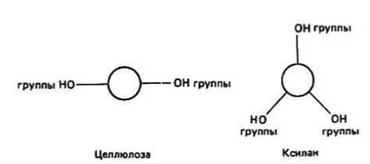
Fig. 31.3.
GLYCOGEN is the reserve polysaccharide of animals, with its highest concentration found in the Liver of mammals. Glycosidic bonds of the a(1 —> 4) type also link the D-glucose chains in glycogen, but its side branches—attached to the main chain via a(1 —> 6) linkages—occur much more frequently than in amylopectin. Unlike starch, glycogen lacks a helical structure. The glycogen molecule is even more branched and, consequently, possesses a more open structure. Because this polysaccharide serves as a rapidly mobilized energy source in animals (utilized more frequently than the corresponding plant reserve substance), it must be readily accessible to enzymes that cleave glucose units from the polymer chain.
Chitin, a structural polysaccharide and the main component of the insect exoskeleton, is formed from chains of slightly modified glucose. If the —OH group at the C2 position of glucose is replaced by a —NH.CO.CH3 (N-acetyl) group, N-acetylglucosamine (NAG) is formed. Polymers of NAG linked by ß(1 —> 4) bonds build a layered structure similar to that of cellulose. However, the interchain Hydrogen Bonds are stronger here due to the involvement of the N-acetyl group. Insects synthesize a multilayered structure in which poly(NAG) sheets alternate with protein layers, resulting in an extremely tough shell. This demonstrates that polysaccharides complexed with Proteins or Peptides are capable of forming a far greater variety of structures than they can on their own (Chap. 35).
HYALURONIC ACID is a polymer featuring alternating ß(1 —> 3) and ß(1 —> 4) linkages between D-NAG monomers and D-glucuronic acid (a modified form of NAG in which the 6CH2OH group is replaced by COOH) in the following sequence: [D-glucuronic acid-(1 —> 3)-NAG-(1 —> 4)]n. Hyaluronic acid is an integral component of Cartilage, bones, and Skin, and is associated with other complex sugars (such as chondroitin sulfate and keratan sulfate) as well as proteins.
Last update: 13/08/2026
Editorial and Educational Adaptation: This material has been compiled based on the primary/original source text. The project team performed an editorial review, corrected technical inaccuracies, structured sections, and adapted the content for an educational format.
What was processed:
- elimination of formatting defects (OCR errors, structural breaks, corrupted characters);
- editorial organization of content;
- standardization of terminology in accordance with academic sources;
- verification of factual statements against the original source text.
All mentions of the author, publication year, and origin of the primary text have been preserved in accordance with the source.