IMMUNOLOGY TEXTBOOK - Mercury Podillya 2013
IMMUNOLOGICAL RESEARCH METHODS
Laser Flow Cytofluorimetry
THE PRINCIPLE OF flow cytometry is based on registering light scatter and fluorescence from each individual Cell within a cell suspension. Based on light scatter analysis (without The Use of Antibodies), the content of lymphocytes, monocytes, and granulocytes in the test sample can be determined. Flow cytometry is performed using Monoclonal Antibodies conjugated with fluorescent Dyes used to stain Blood Cells. Monoclonal antibodies exhibit identical Specificity toward membrane Antigens; therefore, they are grouped and designated with the corresponding cluster of differentiation (CD) number. Thus, utilizing the immunofluorescence technique (direct or indirect), the counts of various lymphocyte subpopulations can be determined.
Among the most commonly used fluorochromes are the following: fluorescein isothiocyanate (FITC), phycoerythrin (PE, RD1), peridinin chlorophyll protein (Per-CP), allophycocyanin (APC), as well as tandem dyes (phycoerythrin-Cy5 and Cy7).
The Cell suspension is pressurized into a flow cell, where, due to the pressure differential between the sample and the sheath fluid, the cells in a laminar liquid flow align in single file (hydrodynamic focusing of a core stream within a sheath). Blood cells cross the focused laser light beam one by one. Light of a specific wavelength excites the molecules of fluorescent dyes bound to various cellular components; simultaneous excitation of several different dyes can occur, enabling the evaluation of multiple cellular parameters at once.
At the moment a cell intersects the laser beam, detectors record:
✵ forward scatter (FSC) (Fig. 3). The forward scatter detector is positioned along the path of the laser beam behind the flow cell and registers laser emission scattered at angles of 2–19°. The intensity of the light scattered at a small angle is proportional to cell size; larger cells scatter light more intensely than smaller ones;
Class="center">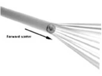
Fig. 3. Laser flow cytometry. Forward scatter (FSC) (explanation in text)
✵ side scatter (SSC) (Fig. 4). Passing through the cell, the laser beam undergoes multiple refractions and scatters in all directions at an angle of 10° or greater. Registration of this emission allows for the Assessment of the cell's internal architecture (Nucleus-to-Cytoplasm ratio, presence of granules and other intracellular inclusions). The combination of side and forward scatter provides insights into overall cell Morphology and allows for the isolation of distinct cell populations (lymphocytes, monocytes, granulocytes) for further analysis;
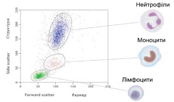
Fig. 4. Laser flow cytometry. Side scatter (SSC) (explanation in text)
✵ fluorescence intensity, which allows for the Determination of the subpopulation COMPOSITION OF THE cell suspension, etc.
The system for recording fluorescent emission consists of a complex of optical filters and photomultipliers, each registering emission within a wavelength range corresponding to the specific fluorochrome. The choice of type and quantity of fluorescent dyes is determined by the specific research objective. The primary types of such dyes include fluorescently labeled monoclonal antibodies (FITC, PE, APC, PerCP, etc.) for identifying membrane and cytoplasmic cellular antigens; dyes for evaluating cell viability (7AAD, PI); nucleic acid-binding fluorophores (DAPI, Hoechst); pH-sensitive fluorophores (Fluo-3); and ion-dependent fluorophores (Indo-1). For instance, CD3 antigens are detected using OKT3, OKT1, and Leu4 monoclonal antibodies, where OK stands for Orto Klon; CD4 antigens using OKT4 and Leu2a; CD8 antigens using OKT8 and Leu3a; and CD2 antigens using OKT11. Monoclonal antibody OKT6 detects CD1 antigens; OKT9 and OKT10 detect antigens on pre-thymic cells, immature and activated T lymphocytes; OKB1 and Leu12 monoclonal antibodies target mature B lymphocytes; OKB2 targets early B lymphocyte forms; and OKM1 and Leu7 target monocytes, granulocytes, and natural killer cells.
The resulting signal is transmitted to a computer, processed, and displayed in the form of various graphs and histograms.
In a flow cytometer equipped with a cell sorting system, the flow cell is mounted on a piezocrystal. When voltage is applied, the crystal, along with the cell, oscillates at a predetermined frequency, causing the fluid jet containing cells to break into individual droplets. Passing through a charging ring, a droplet can acquire a positive or negative charge depending on the cell contained within it. Flying past deflection plates, the droplet with the cell is attracted to them, diverted from the main stream, and collected into a tube. The cell sorting method on a flow cytometer yields cell populations with high purity (up to 99.9% positive cells in the sorted fraction).
Rosette formation assay. Determination of T lymphocytes by spontaneous rosette formation with sheep red blood cells (E-RFC). Thymus-dependent T lymphocytes possess receptors for sheep red blood cells (E-receptors identical to CD2, which is detected by monoclonal antibodies), serving as a specific marker for their identification (E-RFC: Erythrocyte-rosetting cells).
Procedure. 0.1 ml of lymphocyte suspension is added to plastic tubes (2 to 5) along with an equal volume of a 0.5% sheep red blood cell suspension. The erythrocyte-to-lymphocyte ratio should not exceed 50:1. The mixture is incubated in a thermostat at 37°C for 10 minutes. The tubes are then centrifuged at 1000 rpm for 5 minutes and left overnight in a refrigerator at 4°C. Cell counting is performed using a Goryaev chamber. The cell suspension is fixed with glutaraldehyde or acetaldehyde, followed by smear preparation and rosette counting in stained preparations, which allows slides to be stored and reaction results to be analyzed on any subsequent day.
For cell counting in the Goryaev chamber, the cell pellet in tubes retrieved from the refrigerator is gently resuspended using a Pasteur pipette (slowly drawing and expelling the cell suspension several times), and 0.02 ml of a 0.01% acridine orange solution in phosphate buffer is added. This dye produces bright green luminescence when excited by ultraviolet light. After 2–3 minutes, the Goryaev chamber is loaded, and the percentage of E-RFCs is determined by counting 300 lymphocytes under a fluorescence Microscope.
On the day of blood collection for T lymphocytes, a complete blood count must be performed to enable the calculation of absolute T-cell values.
Normal T-cell ranges in healthy Donors: 54.3 ± 0.98%; 979.8 ± 16.8 cells/µl.
Determination of active T lymphocytes forming rosettes with sheep red blood cells (EA-RFC). All preparatory steps are performed as described for E-RFC, except that serum is not added to the incubation medium for EA-RFC determination, and prolonged cold incubation is omitted. Following incubation at 37°C for 10 minutes and subsequent centrifugation at 1000 rpm for 5 minutes, active T lymphocytes are counted as described above.
The content of active T lymphocytes in healthy donors is: 34.6 ± 1.92%, 840 ± 123 cells/ml.
Determination of theophylline-sensitive T cells. In the presence of theophylline, T lymphocytes with suppressor function lose their ability to form E-rosettes. These cells are designated as theophylline-sensitive (TS), the functional counterpart of CD8+ T suppressors. So-called theophylline-resistant (TR) cells in a significant percentage of cases contain a subpopulation of CD4+ T helpers. The normal TR/TS ratio is 2.5–3.5.
Procedure. The Reagents and equipment used were identical to those described above for the determination of active T-lymphocytes. Prior to the assay, a 0.3 M theophylline solution is prepared in distilled Water warmed to 60 °С. The theophylline solution, cooled to room Temperature, is added to the incubation medium (without serum), incubated, centrifuged at 1000 rpm for 5 min, and the cells are counted in the same manner as active T-cells. Two subpopulations are identified: theophylline-sensitive T-cells, i.e., lymphocytes that have lost The ability to form rosettes upon theophylline Treatment, and theophylline-resistant T-cells.
In healthy donors, The ratio of theophylline-sensitive to theophylline-resistant T-cells is 1:3.
Detection of T-lymphocytes forming rosettes with allogeneic and autologous erythrocytes. T-cells that form rosettes with autoerythrocytes are believed to possess killer function and play a key role in autoaggressive mechanisms.
Procedure. The lymphocyte suspension is isolated using the method described above. Human erythrocytes: it is necessary to use Rh-negative group 0(I) erythrocytes. The preparation of erythrocytes is analogous to the method described for sheep erythrocytes. Sheep erythrocytes and human erythrocytes are added simultaneously to the lymphocyte suspension in the same ratios. The lymphocytes that have bound both sheep and human erythrocytes are counted. The reaction with autologous erythrocytes is carried out in the same way as with allogeneic ones.
Determination of B-cells by rosette formation with sheep erythrocytes in the EAC system.
Thymus-independent B-lymphocytes bear specific determinants on their membrane that allow them to differentiate from thymus-dependent cells, i.e., T-lymphocytes. Such determinants include surface (membrane) IgM, receptors for the Fc fragment of IgG, the third Complement component (C3), and the Epstein-Barr virus. The number of lymphocytes bearing IgA, G, E, or D is insignificant (IgA — 1–5 %, IgD and IgE — 2–4 %). The method of detecting B-cells based on their ability to form rosettes with sheep erythrocytes coated with antibodies in a complement-containing medium is applied. Such erythrocytes label the Fc and C3 receptors of B-lymphocytes.
The instruments and reagents are the same as for the T-lymphocyte determination method. The lymphocyte suspension is prepared in a similar manner.
Procedure. The antiserum containing antibodies against erythrocytes is prepared by immunizing rabbits with sheep or bovine red blood cells. A 3-5 mL volume of a 50% erythrocyte suspension is injected into the marginal ear vein of a rabbit. Blood is collected on days 4–6 to obtain the serum, which at this stage (at the peak of the Immune Response) predominantly contains IgM. The best results are achieved using the gamma-globulin fraction of the serum, isolated by salting out in a saturated ammonium sulfate or rivanol solution. The antiserum is then inactivated, and its hemolytic and agglutination titers are determined. Alternatively, ready-to-use commercial rabbit hemolytic serum can be utilized.
Complement. Fresh mouse sera serve as the source of complement. Outbred mice are decapitated, the blood is drained into a test tube to obtain serum, which is subsequently absorbed with a pool of human erythrocytes, and complement activity is determined using a hemolytic system.
Erythrocyte sensitization. Equal volumes of a 1% suspension of sheep (or bovine) erythrocytes and the antiserum specific to THE RED BLOOD cell species used in the assay (or rabbit hemolytic serum) are mixed at a subagglutinating dilution. The mixture is incubated for 40 min at 37 °C with gentle shaking every 10 min. Following incubation, the erythrocytes are washed three times with phosphate buffer by centrifuging at 1500 rpm for up to 10 min. The supernatant is discarded, and the pellet is resuspended in the initial volume of phosphate buffer plus an equal volume of absorbed mouse serum containing complement at a 1:10 dilution. The mixture is placed in an incubator at 37 °C for 30 min. The erythrocytes are then washed three times again. During these washes, centrifugation is performed gently at 1000 rpm for 5 min to prevent red blood cell agglutination. A 0.5% erythrocyte suspension is prepared and examined under a microscope. If agglutination is present, the suspension is unsuitable for use. The prepared antibody- and complement-loaded erythrocytes (EAC) can be stored in a refrigerator (4 °C) for 4–5 days.
Determination of EAC-rosetting lymphocytes. Add 0.1 mL of the lymphocyte suspension to 0.1 mL of the sensitized erythrocyte suspension. The optimal erythrocyte-to-lymphocyte ratio is 20:1. The mixture is incubated for 45 min at 37 °C, after which the tubes are placed on ice. B-cell enumeration is performed as described above.
The normal absolute count of B lymphocytes is 0.28 - 0.31x10⁶ /L.
Clinical significance. An elevated absolute B-lymphocyte count is observed in: acute bacterial, fungal, and parasitic infections, AIDS (early stage), chronic Liver diseases (cirrhosis, Viral Hepatitis), autoimmune disorders (rheumatoid Arthritis, SLE, rheumatic fever, collagenoses), sarcoidosis, cystic fibrosis, Crohn's disease, Waldenström's macroglobulinemia, monoclonal gammopathy, infectious mononucleosis, chronic Lymphocytic Leukemia, and during the acute phase of reinfection.
A decreased absolute B-lymphocyte count is observed in: physiological hypogammaglobulinemia in infants (at 3–5 months of age), congenital hypogammaglobulinemia or agammaglobulinemia, immune system neoplasms, treatment with cytostatics and immunosuppressants, post-splenectomy states, and humoral immunodeficiency.
Last update: 13/08/2026
Editorial and Educational Adaptation: This material has been compiled based on the primary/original source text. The project team performed an editorial review, corrected technical inaccuracies, structured sections, and adapted the content for an educational format.
What was processed:
- elimination of formatting defects (OCR errors, structural breaks, corrupted characters);
- editorial organization of content;
- standardization of terminology in accordance with academic sources;
- verification of factual statements against the original source text.
All mentions of the author, publication year, and origin of the primary text have been preserved in accordance with the source.