Fundamentals of Molecular Biology - V.I. Rezyapkin 2009
Replication
Telomere Synthesis
Somatic Cells can divide a limited number of times; for example, embryonic cells can divide up to 80 times, whereas cells from a 70-year-old human can only divide 20 to 30 times. This finite capacity for Cell Division is known as the Hayflick limit.
In some cases, cells are able to overcome the Hayflick limit and begin dividing indefinitely, a phenomenon referred to as cell immortalization.
After 50 divisions, the total mass of the progeny from a single cell could reach 20,000,000 tons.
The limitation on the number of cell divisions is determined by the presence of specialized structures at the ends of Chromosomes known as telomeres. Telomeres consist of short, repeating DNA sequences. In many species, these are represented by the highly conserved TTAGGG repeats. Telomeric DNA is non-coding. In humans, the length of telomeres can range from 2,000 to 20,000 bp. As an Organism develops, telomere length decreases. Cells with shorter telomeres have a reduced division potential. The shortening of telomeres during cell division is associated with the end-Replication problem of linear chromosomes. This incomplete replication occurs because An RNA primer is required during replication, and DNA Synthesis proceeds in the 5’→3’ direction (Fig. 2.14).
Class="center">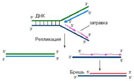
Fig. 2.14. Incomplete Chromosome replication
The enzyme responsible for lengthening telomeres is telomerase. However, at a certain stage of development, the telomerase Gene is switched off. As a result, telomeres shorten with each division, leading to impaired chromosomal function, given that telomeres protect genetic material from degradation and are responsible for proper chromosome positioning and functioning within The Cell. It is worth noting that telomerase genes are deactivated only in sexually reproducing organisms, not in those that reproduce vegetatively.
Telomerase is an ribonucleoprotein enzyme. The RNA component of telomerase contains a trinucleotide complementary to a specific region of the telomere repeat sequence. Their complementary interaction (Fig. 2.15) forms a template region that the enzyme uses to synthesize a new telomere fragment. Upon completion of this fragment synthesis, the RNA component of telomerase translocates toward the 3' end of the newly synthesized chain. Telomerase is then ready to resume DNA synthesis. This cyclic process is repeated multiple times until the Synthesis of the corresponding DNA strand is complete.
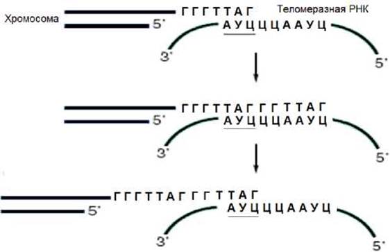
Fig. 2.15. Mechanism of telomerase action
After telomerase completes the synthesis of the DNA strand in the 3’ direction, DNA polymerase and other Proteins facilitate the synthesis of the complementary strand (Fig. 2.15). This is how telomeres are formed.
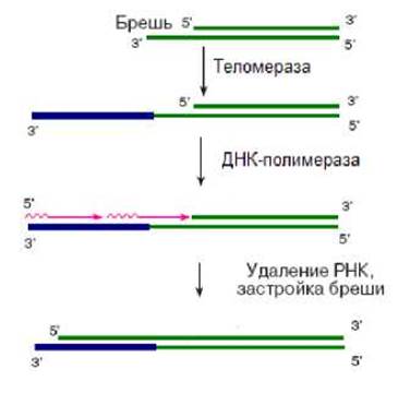
Fig. 2.15. Telomere formation
Telomerase RNA is encoded by genes, and its length varies across different organisms: in Protozoa, it is 150–200 NUCLEOTIDES; in humans and mice, 450 nucleotides; and in Yeast, 1,300 nucleotides.
The template region of the telomerase RNA is located 50 nucleotides away from its 5' end. In mouse telomerase, the RNA template region is 8 nucleotides long, compared to 11 in humans. Nevertheless, both sequences encode the exact same telomeric repeating unit.
In Germ Cells, the telomerase gene is expressed, which results in telomerase activity and telomere synthesis. In somatic cells, however, the telomerase gene is turned off, and no telomerase activity is detected.
In Cancer cells, the telomerase gene is activated, resulting in sufficiently high levels of telomerase activity and subsequent telomere synthesis. Although shorter than those in embryonic cells, telomeres in cancer cells remain stable.
The Use of antisense RNAs directed against the RNA component of telomerase caused the death of HeLa tumor cells. These experiments highlight the crucial role of telomerase in the malignant transformation of normal cells. At the same time, knockout mice lacking the gene encoding the telomerase RNA component have been generated. Although these mice lacked telomerase activity, they remained viable for up to six generations. Furthermore, cell lines derived from these mice underwent immortalization, were transformed by Viral Oncogenes, and became tumorigenic in nude mice. Cells from the fourth-generation mice lacked telomeric repeats and exhibited various abnormalities. These findings confirm that telomerase is essential for maintaining telomeres, but they challenge its direct role in cell immortalization and malignant transformation.
To sum up, it should be noted that the replication of Genetic information is a highly complex biological process involving numerous factors.
Last update: 12/08/2026
Editorial and Educational Adaptation: This material has been compiled based on the primary/original source text. The project team performed an editorial review, corrected technical inaccuracies, structured sections, and adapted the content for an educational format.
What was processed:
- elimination of formatting defects (OCR errors, structural breaks, corrupted characters);
- editorial organization of content;
- standardization of terminology in accordance with academic sources;
- verification of factual statements against the original source text.
All mentions of the author, publication year, and origin of the primary text have been preserved in accordance with the source.