Molecular Biotechnology: Principles and Applications - Glick, B., & Pasternak, J. 2002
Fundamentals of Molecular Biotechnology
Recombinant DNA Technology
Vectors for Cloning Large DNA Fragments
Bacteriophage λ-Based Vectors Plasmid Vectors can be used to clone DNA fragments up to 10 kbp in length. However, constructing Genomic Libraries oft
en requires working with larger fragments. To this end, vectors based on the E. coli bacteriophage λ were developed.
Following The entry of phage λ into an E. coli Cell, infection can proceed via one of two pathways. Under the lytic cycle, the phage rapidly multiplies, and within approximately 20 min the host cell is destroyed (lysed), releasing up to 100 new phage particles. Alternatively, the phage DNA integrates into the E. coli chromosome as a prophage, replicating alongside normal bacterial genes (a state known as Lysogeny). However, under nutrient deprivation or other adverse conditions, the integrated phage DNA is excised, triggering the lytic developmental pathway. The DNA genome of phage λ is approximately 50 kbp in size, a significant portion of which (about 20 kbp) is non-essential for phage propagation and merely governs its integration into the host DNA. This led to the concept that this region could be replaced with a foreign DNA fragment of equivalent size. The resulting recombinant molecule would then replicate within The Cell as the DNA of a "recombinant" phage λ committed to the lytic pathway.
To understand how the phage λ vector system Functions, it is necessary to examine the molecular aspects of the lytic cycle. An infectious phage particle consists of a protein HEAD enclosing tightly packaged DNA approximately 50 kbp long, and a tail with protruding thin protein filaments (fibrils). Head and tail assembly, as well as DNA packaging, are precisely coordinated. The DNA of phage λ is a linear double-stranded molecule of 50 kbp with single-stranded 5' overhangs of 12 NUCLEOTIDES. These are termed cohesive (cos) ends because they are mutually complementary and capable of base-pairing. Once the phage DNA enters the E. coli cell, the cos ends join to form a circular molecule. Early in the lytic cycle, Replication of this circular DNA molecule generates a linear concatemer consisting of multiple 50-kbp segments (Fig. 4.16, A). Each such segment is packaged into a protein head, which subsequently attaches to a preassembled tail to yield a new phage particle (Fig. 4.16, B). Packaging DNA molecules shorter than 38 kbp yields non-infectious phage particles, whereas fragments longer than 52 kbp cannot fit into the head. The 50-kbp segments within the linear DNA molecule are separated by cos sites, and it is precisely at these sites that the molecule is cleaved as each successive segment fills a head. This Cleavage is catalyzed by an enzyme located at the head vertex.
Studies on phage λ assembly led to The Development of an in vitro DNA packaging system that generates infectious phage particles. By mixing purified empty heads, phage DNA, and assembled tails in a test tube, infectious phage particles can be successfully reconstituted.
One of many λ cloning vectors features two BamHI restriction sites flanking a 20-kbp region. Digestion of purified phage DNA with BamHI produces three fragments: the left arm (region L), which contains Genetic information for the phage head and tail; the right arm (region R), which manages METABOLISM/36.html">DNA replication and lysis; and the central segment, which harbors genes responsible for integration and excision (the I/E segment, derived from integration/excision). The researcher's objective is to replace this central region with a desired DNA sequence approximately 20 kbp in length (Fig. 4.17). The DNA to be cloned is likewise digested with BamHI, and fragments ranging from 15 to 20 kbp are isolated. Both preparations—phage DNA and foreign DNA—are combined and treated with T4 DNA ligase, followed by The addition of empty heads and preassembled tails. DNA fragments of 50 kbp are packaged into heads, tails are attached, and infectious phage particles are formed. Fragments that are larger (>52 kbp) or smaller (<38 kbp) cannot be packaged. The recombinant phage λ can only propagate in E. coli strains that do not support the growth of phage λ with an intact I/E region. To maintain the recombinant phage λ, it is periodically passaged on a fresh E. coli culture.
Class="center">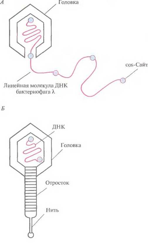
Fig. 4.16. Lytic Pathway of bacteriophage λ development. A. Replication of circular bacteriophage λ DNA generates a linear molecule composed of repeating segments approximately 50 kbp in length. Each of these segments represents a full-sized phage genome. B. The phage head accommodates one such segment, after which the preassembled tail attaches to the head.

Fig. 4.17. Bacteriophage λ-based cloning system. The phage DNA contains two BamHI sites flanking its I/E segment. The DNA to be cloned is digested with BamHI, and the resulting fragments are size-fractionated to isolate those ranging from 15 to 20 kb in length. The phage DNA is treated with the same restriction enzyme. Both DNA preparations are mixed and incubated with T4 DNA ligase. The ligation mixture contains A wide variety of DNA combinations, including 1) reconstituted λ phage DNA, and 2) recombinant molecules consisting of the R and L Regions of the phage DNA and a ~20 kb cloned DNA insert that has replaced the I/E region of the phage genome. These recombinant molecules are packaged into bacteriophage λ heads in vitro, and upon the addition of tails, infectious phage particles are formed. In E. coli Cells infected with the recombinant phage, whose chromosome carries an integrated bacteriophage P2 DNA, only DNA molecules composed of the R and L phage regions and a ~20 kb cloned insert can replicate and form infectious particles.
Library screening for λ-phage-based systems can be carried out using DNA probes or immunological Methods. Lysis zones (plaques) are transferred onto a filter and tested accordingly. When DNA Hybridization is used, phage Proteins are first removed, after which the DNA is denatured and fixed on the filter. In immunological testing, proteins encoded by the cloned genes are transferred and fixed on the filter along with the plaque. By matching the positive spots on the filter with the plaques on the original Agar plate, positive plaques are picked and subcultured. These subcultures serve as a source of recombinant Bacteriophages that can be individually propagated in E. coli.
Cosmids
Vectors known as cosmids can accommodate up to 40 kb of foreign DNA while being actively amplified in E. coli like Plasmids. Cosmids combine the features of plasmid vectors and λ phage-based vectors. For instance, the widely used cosmid pLFR-5 (approximately 6 kb) contains two λ phage cos sites separated by a ScaI restriction site, a polylinker with six unique restriction sites (HindIII, PstI, SalI, BamHI, SmaI, and EcoRI), a DNA replication origin (ori), and a tetracycline resistance Gene (Tetr). This cosmid can accept foreign DNA inserts up to 40 kb in length (Fig. 4.18). DNA fragments of approximately 40 kb intended for cloning are purified by sucrose density gradient centrifugation from partial BamHI digests of donor DNA (Fig. 4.18), whereas pLFR-5 is digested first with ScaI and then with BamHI. The DNA preparations are mixed and ligated. Ligation products containing a 40 kb insert have a total size close to 50 kb and can therefore be packaged in vitro into λ phage heads. Reassociated pLFR-5 molecules lacking inserts will not be packaged. Following phage particle assembly, they are used to infect E. coli (Fig. 4.18).

Fig. 4.18. Cloning using a cosmid vector. The cosmid features a replication origin (ori) ensuring its maintenance in E. coli as a plasmid; two intact cos ends separated by a unique ScaI site; a BamHI site near one of the cos ends; and a tetracycline resistance gene (Tetr). The DNA targeted for cloning is digested with BamHI and size-fractionated to isolate molecules of approximately 40 kb. The plasmid DNA is digested with ScaI and BamHI. Both DNA preparations are mixed and treated with T4 DNA ligase. Some of the hybrid molecules formed after ligation contain an insert of about 40 kb, yielding a total length of approximately 50 kb. These molecules are packaged in vitro into bacteriophage λ heads, tails are subsequently attached to the heads, and infectious particles are formed. Upon infection of E. coli with this "phage," a linear DNA molecule with cos ends is released into the bacterial cell; these ends pair with each other, host cell DNA ligase seals the single-stranded nicks, and the resulting circular molecule persists in the host cell as an autonomously replicating unit. Transformed cells can be identified by their resistance to tetracycline.
Once inside the bacterial cell, the linear pLFR-5 molecule with its insert circularizes via the base-pairing of its cos sites. In this stable configuration, it can persist within the cell for a long time and replicate as a hybrid plasmid, as it contains all the necessary elements. Furthermore, the tetracycline resistance gene enables the growth of colonies carrying this cosmid on media containing the antibiotic, whereas non-transformed cells perish. Other λ-phage-based cosmid vectors also exist.
Cosmids offer a major advantage over plasmids: they can accommodate much larger DNA fragments, meaning that constructing a genomic library requires fewer clones and less time for screening.
Vector systems for cloning very large DNA fragments. Vector systems capable of harboring large inserts (>100 kb) are invaluable for analyzing complex Eukaryotic Genomes. Such vectors are indispensable, for instance, in human Genome Mapping or gene identification. Unlike libraries with small inserts, a genomic library with large inserts is far more likely to represent the entire genetic material of an Organism. Moreover, this approach reduces the number of clones that need to be maintained and increases the probability that any given gene will be present within its specific clone. To clone DNA fragments ranging from 100 to 300 kb in length, a low-copy-number plasmid vector based on bacteriophage P1 was constructed—a chimeric construct known as a P1-derived artificial chromosome. Additionally, a highly stable vector capable of accepting inserts from 150 to 300 kb was developed on The basis of the E. coli F-plasmid (F factor, or fertility factor), which is present in the cell as one or two copies, incorporating the lacZ' Selection system of pUC vectors. This construct is designated as a bacterial artificial chromosome (BAC).
Last update: 11/08/2026
Editorial and Educational Adaptation: This material has been compiled based on the primary/original source text. The project team performed an editorial review, corrected technical inaccuracies, structured sections, and adapted the content for an educational format.
What was processed:
- elimination of formatting defects (OCR errors, structural breaks, corrupted characters);
- editorial organization of content;
- standardization of terminology in accordance with academic sources;
- verification of factual statements against the original source text.
All mentions of the author, publication year, and origin of the primary text have been preserved in accordance with the source.