Orthopedics - Oleksa A.P. 2006
Joint Pathophysiology
Developmental Defects and Congenital Skeletal Anomalies
Paget's Disease
This disease was first described by Sir James Paget in 1877 under the name osteitis deformans, as he believed it to be of inflammatory origin. However, in 1924, T. Stenholm demonstrated the dystrophic Nature of the condition and introduced the term fibrous osteodystrophy.
The core pathology involves the remodeling of Bone tissue characterized by alternating cycles of resorption and new Bone Formation, resulting in a mosaic-like Bone Structure.
The exact Etiology of Paget's Disease remains unclear, although several hypotheses have been proposed: a congenital inherited recessive defect in The Biosynthesis of bone Connective Tissue, as well as endocrine, autoimmune, vascular, neoplastic, dysplastic, and even viral factors—supported by the detection of virus-like inclusions within the nuclei of osteoclasts from affected sites.
High levels of alkaline phosphatase activity and hydroxyprolinuria indicate that severe bone Metabolic Disorders lie at the ROOT of the disease Pathogenesis.
Monostotic lesions of the Tibia or Femur are most common, whereas involvement of other bones, including the spine and Skull, is extremely rare. Polyostotic forms may affect several bones simultaneously, but they are never generalized as is the case in hyperparathyroid osteodystrophy.
The condition typically manifests in individuals over 40 years of age, more frequently in women, although some authors distinguish a juvenile form, while others classify such presentations as hyperphosphatasia.
The onset of the disease is difficult to determine, as patients only gradually begin to experience mild pain in the affected bone, usually seeking orthopedic consultation when a Varus deformity of the tibia or another bone develops, or secondary coxarthrosis or protrusion of the femoral HEAD occurs.
Long tubular bones appear thickened and bowed under physiological loads. Local hyperthermia can be detected, caused by hypervascularization of the affected area and overlying subcutaneous vessels.
Blood Laboratory tests reveal no abnormalities other than elevated alkaline phosphatase levels. Urinalysis shows only increased hydroxyprolinuria.
The Diagnosis of Paget's disease is primarily based on radiological findings. Radiographs in two projections reveal alterations in the internal trabecular architecture of the affected bone, presenting as a coarse-meshed restructuring of the bone pattern. Bone trabeculae are thickened, coarse, and aligned along lines of mechanical stress, interspersed with irregular radiolucent areas (Fig. 73).
The remodeling process simultaneously involves the fibrous-structured cortical layer. In the late Stages of the disease, the boundary between the cortex and the cancellous bone disappears. The bone acquires a characteristic appearance: due to alternating areas of sclerosis and rarefaction, it thickens and bows while retaining its overall contour (Fig. 74). Concurrently with bone thickening, the medullary cavity progressively narrows and eventually disappears as the Bone Marrow is replaced by fibrous tissue.
Weight-bearing on a bowed bone induces structural remodeling along its convex side, leading to The formation of Looser's zones (Fig. 75), which may subsequently progress to a complete transverse fracture.
Paget's disease may affect one or several vertebrae. The pathological process typically localizes to the vertebral body or simultaneously to its arches, most frequently involving several adjacent vertebrae. The entire spinal Column is never affected. The condition may localize in the lumbar or thoracic spine, and very rarely in the cervical region. Vertebral bodies increase in volume, and their lateral and anterior surfaces become flattened. Occasionally, vertebral bodies undergo compression, becoming flattened and projecting anteriorly relative to normal vertebrae, or posteriorly, thereby narrowing the spinal canal and intervertebral foramina.
Radiological examination reveals radiolucent areas within the vertebral bodies interspersed with coarse trabecular bundles, and occasionally areas resembling fine-grained pumice. The articular interface between the vertebra and the thinned intervertebral disc is irregular and notched.
Class="center">
Fig. 73. Affected femur in Paget's disease in a 55-year-old female, shown in two projections.
It should be noted that Paget's disease can affect the cranial bones and the skull base, leading to various neurological disorders (such as trigeminal or optic neuritis) as well as disturbances in CEREBROSPINAL FLUID dynamics.
Treatment for Paget's disease is primarily conservative. Given the various etiological hypotheses, corticosteroids, androgens, anabolic Hormones, cytostatics, and cytotoxic agents are prescribed, alongside multivitamin therapy and general supportive care. Local radiotherapy is occasionally used.
Surgical intervention is indicated for pathological fractures of long bones, arthrosis, and spinal canal or intervertebral foramina stenosis when radicular pain is refractory to conservative management.
Patients with Paget's disease must be placed under dispensary observation and evaluated twice a year, as pathological bone lesions can be complicated by benign or malignant neoplasms. Sarcomas occur in 10% of cases of Paget's disease, histologically presenting as osteogenic pleomorphic Cell sarcomas, chondrosarcomas, fibrosarcomas, and reticulosarcomas.
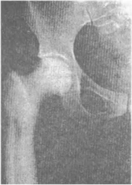
Fig. 74. Thickening of the upper third of the femur with signs of sclerosis and narrowing of the medullary cavity in Paget's disease.
If a tumor process is detected or malignancy is suspected, patients are referred to an onco-osteologist.
Last update: 10/08/2026
Editorial and Educational Adaptation: This material has been compiled based on the primary/original source text. The project team performed an editorial review, corrected technical inaccuracies, structured sections, and adapted the content for an educational format.
What was processed:
- elimination of formatting defects (OCR errors, structural breaks, corrupted characters);
- editorial organization of content;
- standardization of terminology in accordance with academic sources;
- verification of factual statements against the original source text.
All mentions of the author, publication year, and origin of the primary text have been preserved in accordance with the source.