Orthopedics - Oleksa A.P. 2006
Spine conditions causing pain
Diseases of the intervertebral joints
The zygapophyseal (facet) joints are of the synovial type, featuring articular Cartilage and a Joint Capsule. They bear nearly 20% of the spinal load, restrict the range of motion in all planes, and thus provide more than 50% of spinal stability. Load on these joints increases significantly during spinal hyperextension.
Facet joint instability eventually leads to intervertebral disc instability, ligamentous hypertrophy, osteoarthritis, and osteophyte formation. Osteophytes of the superior articular process progressively enlarge, narrowing the intervertebral foramen, while those of the inferior articular process narrow the spinal canal.
Ghormley of the Mayo Clinic (Ghormley R.K., 1933) described the so-called facet syndrome. To confirm that back pain can stem from pathological processes in this joint, Mooney and Robertson (Mooney V., Robertson J., 1976) injected physiological saline or cortisone into the facet joints of healthy individuals and proved this hypothesis.
However, other researchers have expressed doubt regarding the existence of such a syndrome, as the injection of procaine solution did not relieve pain in all patients.
Ghormley, Mooney, and other authors point to the following signs of facet joint pathology:
1. Pain occurs suddenly during sudden bending or twisting of the spine.
2. Typically, the pain is unilateral in the lumbosacral region, frequently radiating to the posterior, lateral, or anterior thigh, or the hip joint.
3. The patient experiences a constant, deep, dull ache, almost invariably above the knee. This pain frequently awakens the patient from Sleep.
4. Pain is aggravated by spinal extension and rotation.
5. Pain during hip joint movements can be so intense that patients and physicians alike often attribute it to pathology within the hip joint itself.
6. Physical examination reveals normal Muscle strength, sensation, and Reflexes.
7. The facet joint is tender to Palpation.
8. Intra-articular injection of a procaine or hydrocortisone solution relieves the pain.
To pinpoint the local source of pain, a Differential Diagnosis must be performed during the patient's examination.
When a nerve ROOT is compressed by a displaced fragment of a damaged intervertebral disc, narrowing of the intervertebral foramen by an osteophyte, or As a result of deforming osteoarthritis (Fig. 297), the pain is restricted to the level of the affected monosegment, accompanied by muscle weakness, paresthesia, and even loss of reflexes.
Narrowing of the spinal canal to 10 mm or more may be caused by osteophytes, spondylolisthesis, a displaced intervertebral disc fragment, or ligamentous hypertrophy, and can thus cause a burning pain, typically bilateral in the back and lower extremities. This pain is sharply exacerbated by spinal hyperextension and alleviated by trunk flexion. Consequently, patients find relief in a sitting position, while riding a bicycle, walking uphill, and so on.
Class="center">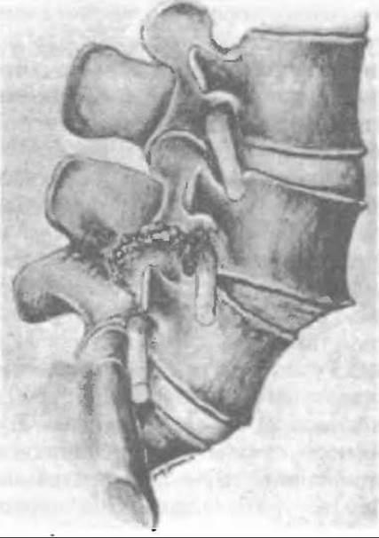
Fig. 297. Nerve root compression in the intervertebral foramen due to degenerative osteoarthritis and spondylolisthesis.
In angiopathies, patients experience unilateral or bilateral back pain caused by physical muscle overload, which occurs during prolonged walking, manual labor, etc.
To clarify the diagnosis, functional X-ray imaging, computed tomography or Magnetic Resonance imaging, radioisotope scanning (technetium-99m), and Laboratory tests are performed.
Treatment depends on the specific pathological changes identified in the facet joint.
If an inflammatory process is confirmed by radioisotope scanning, antibacterial therapy should be prescribed. For acute pain, an intra-articular injection of a procaine solution combined with a corticosteroid preparation provides short-term pain relief in 50–70% of patients and longer-term relief (up to a year or more) in 15–20% of patients. Spinal immobilization with a brace, anti-inflammatory and desensitizing drugs, and physical/balneotherapy are prescribed. Such treatment is quite effective.
When degenerative-dystrophic changes with osteophyte proliferation are detected in the joint, conservative therapy is often ineffective because nerve root compression remains unrelieved.
In the 1970s and 1980s, rhizotomy, proposed by Shealy in 1970 (Shealy C.N., 1975), was utilized. However, the outcomes of this Procedure were unsatisfactory because electrocoagulation led to joint denervation and soft tissue Burns; therefore, rhizotomy is no longer used today.
Currently, the most common approach is a radical procedure consisting of decompressive hemilaminectomy with widening of the intervertebral foramen and facet joint arthrodesis (Figs. 298, 299).
According to Markwalder et al. (Markwalder T.M., Dubach R., Braun M., 1995), 75% of operated patients experienced good outcomes from this surgery. The most successful Procedures were those performed within a single segment, as no pain recurrence was observed following arthrodesis.
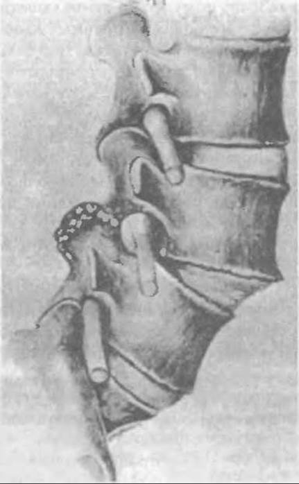
Fig. 298. Decompression of the nerve root via resection of marginal outgrowths of the intervertebral joint.
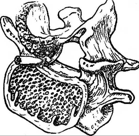
Fig. 299. Decompression of the nerve root via resection of the Base of the superior articular process.
Last update: 10/08/2026
Editorial and Educational Adaptation: This material has been compiled based on the primary/original source text. The project team performed an editorial review, corrected technical inaccuracies, structured sections, and adapted the content for an educational format.
What was processed:
- elimination of formatting defects (OCR errors, structural breaks, corrupted characters);
- editorial organization of content;
- standardization of terminology in accordance with academic sources;
- verification of factual statements against the original source text.
All mentions of the author, publication year, and origin of the primary text have been preserved in accordance with the source.