Cytology, General Histology and Embryology - V. K. Napkhanyuk 2002
Microscopes. Microscopy Technique
Microscopy Technique
Polarization Microscopy
Microscopic examinations in polarized light are performed to study the optical anisotropy of objects. Birefringence in biological specimens occurs due to the longitudinal orientation of rod-like submicroscopic particles, mechanical stress, and other factors.
Polarized light Cell/15.html">Microscopy is used to determine particle orientation, direction of deformation, magnitude of birefringence (the difference between the refractive indices of ordinary and extraordinary rays), and the optical path difference between ordinary and extraordinary rays within the specimen.
The Study of anisotropic objects utilizes a polarizing microscope, which differs from a standard Light Microscope (Fig. 5). In this microscope, a polarizer is placed before the condenser. A compensator and an analyzer are mounted after the specimen and objective to enable comprehensive analysis of birefringence in the sample.
The specimen image is viewed through the eyepiece. Examinations are conducted with the polarizer and analyzer in parallel and crossed positions. In the latter case, in the absence of a specimen, the field of view appears dark. Bright or colored structural elements become visible in the specimen, their appearance depending on the sample's absorption relative to the plane of polarization and the magnitude of birefringence.
Class="center">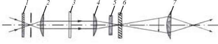
Fig. 5. Principal optical scheme of a polarizing microscope:
1 — polarizer; 2 — condenser; 3 — specimen; 4 — objective; 5 — compensator; 6 — analyzer; 7 — eyepiece
When performing examinations in polarized light, it is essential that the rotating stage is equipped with a graduated scale (limbus), and that the condenser and objective lenses are free from internal strain.
An artifact (artefactum) is an artificial Structure that appears in the test specimen during preparation for study and may lead to unreliable results.
Artifacts can be crude and very simple, making them easy to recognize, but they can also be subtle and extremely difficult to identify.
Examples of simple artifacts include air bubbles trapped in the preparation when the coverslip is applied.
More complex artifacts include cellular deformation, as well as the appearance of cavities or gaps between surrounding tissue layers caused by tissue compression during fixation, dehydration, and related Procedures.
1. STRUCTURE OF THE light microscope.
2. Structure of The Optical System of the light microscope.
3. Technique of microscopy using a light microscope.
4. Determination of the resolving power of a light microscope.
5. Ultraviolet Microscopy. Design principles of a microscope for ultraviolet microscopy.
6. Fluorescence Microscopy. Design principles of a microscope for fluorescence microscopy.
7. Phase-contrast microscopy. Design principles of a microscope for phase-contrast microscopy.
8. Dark-field microscopy. Structure of a microscope for dark-field microscopy.
9. Polarizing microscopy. Structure of a polarizing microscope.
10. Electron microscope. Types of electron microscopes and their characteristics.
11. Operating principle of the electron microscope.
12. METABOLISM/2.html">THE CONCEPT OF artifacts.
Sample exam questions
1. Structure of the light microscope.
2. Rules for operating the light microscope.
3. Design principles of the fluorescence microscope.
4. Electron microscope. Operating principle of the electron microscope.
Last update: 10/08/2026
Editorial and Educational Adaptation: This material has been compiled based on the primary/original source text. The project team performed an editorial review, corrected technical inaccuracies, structured sections, and adapted the content for an educational format.
What was processed:
- elimination of formatting defects (OCR errors, structural breaks, corrupted characters);
- editorial organization of content;
- standardization of terminology in accordance with academic sources;
- verification of factual statements against the original source text.
All mentions of the author, publication year, and origin of the primary text have been preserved in accordance with the source.