Medical Radiology - Lazar A.P. 2008
Radiation Examination of the Maxillofacial Region
The primary radiation imaging method used today for the initial Diagnosis of pathologies in the maxillofacial region is radiography. Dental radiographs can be obtained using any X-ray diagnostic unit, with dental X-ray machines being the most convenient (Fig. 298).
Class="center">
Fig. 298. Dental X-ray machine.
Computed tomography (CT) is utilized in dentistry either as a standalone imaging modality or as an additional method following conventional radiography. It is primarily employed to examine the Maxilla, particularly the maxillary sinuses, providing crucial data on the localization and extent of pathological processes. Maxillofacial CT also allows for the Assessment of the mandibular canal topography, provides information on the Structure and Topography of the inner and outer cortical plates of the jaw bones, their relationship with dental roots, and, in children, with tooth germs. Such examinations are extremely informative for detecting abnormalities in the TEMPOROMANDIBULAR JOINT AND maxillary sinuses, as well as for precisely determining the Location and position of impacted and dystopic Teeth. CT has also found application in evaluating the mineral density of cranial bones. The density index of the hard Tissues of the dentofacial system is one of the key parameters characterizing the state of these tissues and their susceptibility or resistance to pathological changes.

Fig. 299. Orthopantomograph.

Fig. 300. Principle of orthopantomographic image formation.
Magnetic Resonance imaging (MRI) and ultrasound are used primarily to examine the soft tissues of the maxillofacial region. Additionally, MR angiography is employed for the non-invasive visualization of Blood Vessels in this area.
Orthopantomography (panoramic tomography, pantomography) is a tomographic X-ray technique that captures an image of a tomographic layer of both the upper and lower jaws on a single film. This Procedure is performed using a specialized X-ray apparatus known as an orthopantomograph (Fig. 299). The operating principle of the orthopantomograph is as follows: an X-ray film housed in a specialized cassette and the X-ray tube rotate synchronously in opposite directions around the patient's HEAD (Fig. 300). The X-ray beam passes through a vertical Diaphragm slit and progressively exposes the entire film as it rotates. The resulting images are called orthopantomograms, which display a flattened, unrolled view of the jaws and teeth (Fig. 301).
The advantages of this method include the speed of image acquisition—providing information on the state of the patient's entire dentofacial system—at a relatively low radiation dose. However, there are drawbacks: the images of the jaws and teeth are subject to distortion and magnification.
Orthopantomography is often the sole radiological method required and sufficient to establish a diagnosis; sometimes, the analysis of orthopantomograms serves as the basis for further examinations using other X-ray techniques, notably targeted intraoral radiography. This method is used to examine a specific tooth, a small group of teeth, or a distinct section of the alveolar process to study the condition of dental hard tissues, interdental septa, the external and internal cortical plates of the jaws, and the periodontal Bone tissue surrounding the teeth anteriorly and posteriorly, as well as to monitor the quality of endodontic Treatment.

Fig. 301. Orthopantomogram.
Because the anatomical details of dental tissues, periodontium, and jaw bone are quite minute, they can only be visualized on high-quality radiographs. Therefore, an X-ray must meet several requirements: it must fully cover the area of interest and exhibit good definition and contrast. Meeting these criteria ensures that all anatomical structures of the teeth and periodontium, along with any pathological changes, are accurately depicted on the radiograph.
To achieve maximum image sharpness, the X-ray film should be placed as close as possible to the object being examined while simultaneously increasing the distance between the object and the X-ray tube. These conditions also ensure the highest degree of dimensional fidelity between the radiographic image and the anatomical object.
Intraoral radiographs are divided into periapical (contact) and occlusal views. For periapical radiographs, the film is placed inside the Oral Cavity and pressed against the alveolar ridge by the patient's finger. For occlusal radiographs, the film is secured by the patient biting down on it. Periapical radiographs are the primary method because they produce the least amount of tooth distortion.
Occlusal projections are mainly used for maxillary teeth. A 7x6 cm film for adults or a 4x5 cm film for children is placed in the Mouth perpendicular to the sagittal plane.
Radiological examination of the dentofacial system is not limited to images of the teeth and alveolar processes alone, but extends to all PARTS OF THE jaws, including the maxillary sinuses and temporomandibular joints. Images of these structures are obtained using extraoral techniques. Extraoral dental images tend to be distorted and lack structural detail, and are therefore used for diagnostic purposes only when intraoral radiography is impractical for some reason. These exposures are taken in straight (anteroposterior/posteroanterior), lateral, axial, and semi-axial projections.
Extraoral radiographs serve as supplementary diagnostic tools. They are most frequently performed following intraoral imaging to investigate bone lesions in the Mandible. Extraoral radiographs are taken using intensifying screens with 13x18 cm and 18x24 cm films.
The Introduction of radiovisiography into dental practice has significantly expanded the capabilities of dental radiography. In radiovisiography, the traditional X-ray film is replaced by a specialized electronic sensor, and the captured image is processed by computer software and displayed on a monitor screen. This technology allows for digital enhancement of dental images (adjusting brightness and contrast), reduces patient radiation exposure, and provides quantitative data on physical properties, such as the density of the examined tissues.
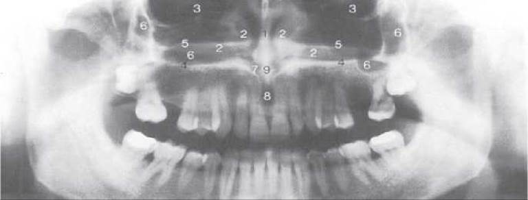
Fig. 302. Anatomical structures on an orthopantomogram: 1 - nasal septum; 2 - Inferior nasal concha; 3 - Orbit; 4 - inferior wall of the Nasal cavity; 5 - horizontal plate of the Palatine bone; 6 - maxillary sinus; 7 - nasal entrance to the incisive canal; 8 - incisive foramen; 9 - anterior nasal spine and nasal crest of the maxilla.
Last update: 08/08/2026
Editorial and Educational Adaptation: This material has been compiled based on the primary/original source text. The project team performed an editorial review, corrected technical inaccuracies, structured sections, and adapted the content for an educational format.
What was processed:
- elimination of formatting defects (OCR errors, structural breaks, corrupted characters);
- editorial organization of content;
- standardization of terminology in accordance with academic sources;
- verification of factual statements against the original source text.
All mentions of the author, publication year, and origin of the primary text have been preserved in accordance with the source.