Medical Radiology - Lazar A.P. 2008
Introduction
History of Radiology
In the late 19th century, scientists across many European countries were conducting experiments with cathode tubes. Cathode radiation was discovered in 1859 by the German mathematician and physicist Julius Plücker. Even before Röntgen, German physicist Philipp Lenard, Riga-born physicist Arthur von Ettingshausen, and Russian physicist Kamensky investigated the rays emanating from a vacuum cathode tube (Crookes tube) when powered by a high-voltage generator (Ruhmkorff coil). They observed fluorescence and photochemical effects, yet failed to fully grasp The Significance of their research.
A native of the Ternopil region (the town of Hrymailiv), physicist Ivan Puluj (1845–1918), who was educated and worked in Austria-Hungary, began publishing articles on cathode radiation in 1877. Puluj and Wilhelm Conrad Röntgen were contemporaries and personal acquaintances; they had studied under the same professor, August Kundt, at the University of Strasbourg, where Puluj earned his doctorate in natural philosophy in 1877. In 1882, he designed a phosphorescent lamp that served as a model for British and German scientists researching cathode rays. Among them was Röntgen, who utilized Puluj's Glass tubes and lamps. From 1884 onward, Ivan Puluj served as a professor and HEAD of the Department of Experimental and Technical Physics at the German Higher Technical School in Prague. In 1892, he published a schematic description of a tube emitting X-rays.
However, the official announcement regarding the discovery of X-rays was made by the German physicist Wilhelm Conrad Röntgen (1845–1923), who had been a professor at the University of Würzburg since 1888 and was elected its rector in 1894. On November 8, 1895, while conducting experiments with a Puluj cathode tube, he noticed a luminescence on a nearby screen coated with barium platinocyanide. Röntgen covered the Crookes tube with black paper, yet the screen continued to glow. Over the next two weeks, virtually locking himself in the laboratory, he investigated The properties of the cathode tube's radiation. He concluded that the unknown rays penetrate bodies depending on their atomic mass, density, and layer thickness; cause ionization; and exhibit biological effects. He established that they do not bend around obstacles but propagate rectilinearly, and shared certain similarities with light (The ability to cast shadows, induce fluorescence, and trigger photochemical reactions). However, he mistakenly hypothesized that the new radiation consisted of longitudinal rather than transverse electromagnetic waves. Röntgen described the Properties of the newly discovered radiation with such remarkable detail in his initial article—published on December 28, 1895, in the Proceedings of the Würzburg Physical-Medical Society—and in two subsequent papers titled On a New Kind of Rays, that almost nothing substantial was added to his findings over the next 17 years. It was not until 1912 that the German physicist Max von Laue, through Interference and diffraction experiments, demonstrated that X-radiation is electromagnetic in nature and differs from light solely by its shorter wavelength.
Class="center">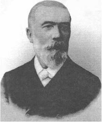
Ivan Puluj
In 1901, Röntgen was awarded the Nobel Prize in Physics; however, he declined the patent for his invention and requested that, upon his death, all his unpublished notes and laboratory journals, which he had kept strictly confidential, be burned.
Immediately after Röntgen's discovery became world-famous, efforts began toward the medical application of the new type of radiation. Initially, X-rays were used to detect bone fractures and foreign bodies. As early as 1896, V. M. Tonkov employed X-rays to study ossification processes and Bone tissue development, while A. I. Lebedev suggested their use in obstetrics and gynecology. Later, with advancements in X-ray technology, The Development of new diagnostic techniques, and the Introduction of artificial contrast media, X-rays came to be widely used for examining the chest Organs, gastrointestinal tract, Kidneys, Urinary System, and more.
In Ukraine, as in other post-Soviet countries, the historically established term "X-rays" (рентгенівське проміння) remains commonly used today, as recommended by the First International Congress of Radiology, whereas abroad the term "X-rays," used by Röntgen himself, is traditionally widespread. Over time, as Ukrainian radiology gradually integrates into international radiological organizations, the terms "X-rays" and "X-ray Examination" will increasingly supersede the older terminology in professional practice.
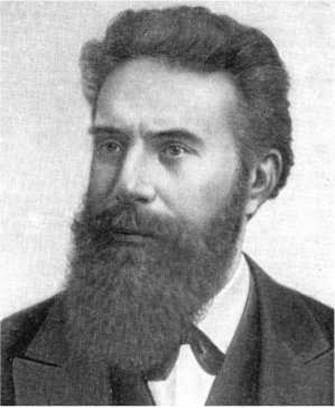
Wilhelm Conrad Röntgen
Last update: 08/08/2026
Editorial and Educational Adaptation: This material has been compiled based on the primary/original source text. The project team performed an editorial review, corrected technical inaccuracies, structured sections, and adapted the content for an educational format.
What was processed:
- elimination of formatting defects (OCR errors, structural breaks, corrupted characters);
- editorial organization of content;
- standardization of terminology in accordance with academic sources;
- verification of factual statements against the original source text.
All mentions of the author, publication year, and origin of the primary text have been preserved in accordance with the source.