Medical Radiology - Lazar A.P. 2008
Radiological examination of bones and joints
The primary method for Radiological examination of Bones and joints is conventional radiography. Radiographs are routinely obtained in two projections—anteroposterior (AP) and lateral—and occasionally supplemented by optimal additional views.
In addition to standard radiography, the imaging toolkit for The Musculoskeletal System includes electroroentgenography (Fig. 28), linear tomography, and contrast-enhanced techniques: angiography for The Vascular System of bones and joints, arthrography for joint cavities, and fistulography for fistulous tracts. Fluoroscopy does not adequately depict Bone Structure or the fine details of pathological processes; therefore, it is rarely used, mainly for the provisional localization of foreign bodies. Linear tomography, by contrast, is useful for identifying small cavities and localized structural alterations.
Recently, linear tomography has been largely superseded by X-ray Computed Tomography (CT), which provides precise localization of pathological lesions, details their internal structure, and measures densitometric density. To optimize image analysis in CT, two primary window settings are typically employed: bone window and soft-tissue window.
The degree of bone mineralization within a specific, limited region of the Skeleton can be assessed using specialized densitometers coupled with computer Processing of X-ray absorption data. Ultrasound densitometry evaluates the acoustic properties (such as ultrasound transmission) of cancellous bone, for instance, in the calcaneal tuberosity, thereby providing an estimate of overall skeletal mineralization.
Magnetic Resonance imaging (MRI) clearly differentiates Muscles, tendons, ligaments, Cartilage, fat, and fluid, making it exceptionally sensitive for detecting soft-tissue and Bone Marrow abnormalities. However, it is less effective for evaluating fine bone architecture and identifying calcifications. Both MRI and CT can be performed even when a limb is immobilized in a plaster cast (Fig. 29).
Class="center">
Fig. 28. Electroroentgenogram of the right ankle region.
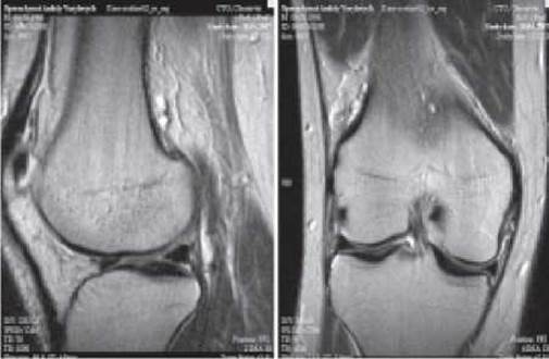
Fig. 29. Magnetic resonance image of the knee (T2-weighted sequence) in the sagittal and frontal planes.
Ultrasound is widely used to evaluate soft-tissue conditions. Because it is free of contraindications, it is frequently chosen for pediatric patients. Common indications for sonography include injuries to joints, tendons, and ligaments, detection of foreign bodies, joint effusions, abscesses, and hematomas, as well as assessment of hip joint instability in developmental Dysplasia. Radionuclide scintigraphy, utilizing technetium-99m-labeled phosphate compounds, allows for whole-body skeletal imaging to detect metabolic bone changes driven by increased osteoblastic activity—such as those seen in osteoblastic tumors and metastases, Osteomyelitis, Arthritis, and post-traumatic or fracture healing. Conversely, purely osteolytic processes, such as osteolytic metastases and multiple myeloma, may yield false-negative results. A normal scintigram effectively rules out malignancy when a sclerotic lesion is identified on a radiograph. Furthermore, scintigraphy offers a distinct advantage in detecting early skeletal abnormalities before they become apparent clinically.
Accordingly, the primary indications for initial osteoscintigraphy are: 1) clinical suspicion of multiple or systemic skeletal lesions; 2) suspected osteomyelitis within the first 10–15 days of onset; and 3) screening for skeletal metastases in patients with a confirmed Diagnosis of Cancer (primarily breast, prostate, thyroid, lung, and renal carcinomas). A limitation of standard planar scintigraphy is the occasional difficulty in accurately localizing pathological foci (for example, differentiating increased radiotracer uptake in the scapula from that in the posterior Ribs, or in vertebral bodies versus posterior spinal elements); however, SPECT successfully overcomes this drawback.
Last update: 08/08/2026
Editorial and Educational Adaptation: This material has been compiled based on the primary/original source text. The project team performed an editorial review, corrected technical inaccuracies, structured sections, and adapted the content for an educational format.
What was processed:
- elimination of formatting defects (OCR errors, structural breaks, corrupted characters);
- editorial organization of content;
- standardization of terminology in accordance with academic sources;
- verification of factual statements against the original source text.
All mentions of the author, publication year, and origin of the primary text have been preserved in accordance with the source.