Human Anatomy - H. I. Koliadenko 2009
Musculoskeletal System
Skeleton of the Upper Limb
Structure and Joints of the Bones of the Upper Limb
The Pectoral Girdle consists of the clavicle and the scapula.
The clavicle is a long S-shaped bone featuring a thickened sternal end and a flattened acromial end. It lies almost horizontally, curving anteriorly, and laterally curves posteriorly (Fig. 28). On its inferior surface, there is an articular facet to which ligaments are attached, connecting it to the first rib and the coracoid process of the scapula. The superior surface of the clavicle is smooth, while the inferior surface is rough.
Class="center">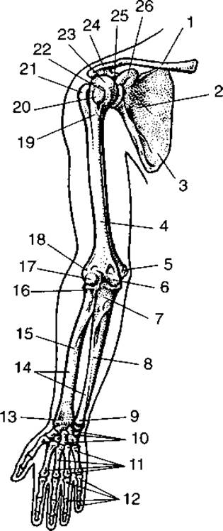
Fig. 28. BONES OF THE upper limb:
1 — clavicle; 2 — subscapular fossa; 3 — scapula; 4 — humerus; 5 — medial epicondyle; 6 — trochlea of the humerus; 7 — tuberosity of the ulna; 8 — ulna; 9 — HEAD of the ulna; 10 — Carpal Bones; 11 — Metacarpal bones; 12 — Phalanges of the fingers; 13 — styloid process; 14 — interosseous borders; 15 — radius; 16 — head of the radius; 17 — capitulum of the humerus; 18 — lateral epicondyle; 19 — surgical neck; 20 — lesser tubercle; 21 — greater tubercle; 22 — anatomical neck; 23 — head of the humerus; 24 — acromion; 25 — glenoid cavity of the scapula; 26 — coracoid process
The scapula is a triangular bone (Fig. 29) with three borders—medial, lateral, and superior—and three angles—superior, inferior, and lateral. The lateral angle is thickened and features the glenoid cavity for articulation with the head of the humerus. Superior to the articular surface lies the coracoid process. On the posterior (dorsal) surface of the scapula, in its upper third, is a prominent bone ridge known as the spine of the scapula. It extends transversely across the scapula from its superior angle to the lateral angle, where it terminates in a flattened process called the acromion, which bears an articular facet for articulation with the clavicle. Superior to the scapular spine is the supraspinous fossa, and inferior to it is the infraspinous fossa.

Fig. 29. Right scapula (posterior view):
1 — superior angle; 2 — supraspinous fossa; 3 — spine of the scapula; 4 — superior border; 5 — coracoid process; 6 — acromion; 7 — lateral angle; 8 — infraspinous fossa; 9 — lateral border; 10 — inferior angle; 11 — medial border
On the costal (ventral) surface of the scapula is a depression called the subscapular fossa, which serves as the origin for the Muscle of the same name.
The clavicular notch of the Sternum and the sternal end of the clavicle form the sternoclavicular joint.
Due to the lack of congruence between these bones, an articular disc is interposed between their articular surfaces, dividing the joint cavity into two compartments. The joint is reinforced by two ligaments: the sternoclavicular and interclavicular ligaments. Additionally, the costoclavicular ligament spans between the first rib and the clavicle. The sternoclavicular joint allows for movements along the sagittal axis (elevation and depression) and the vertical axis (protraction and retraction), as well as axial rotation around the longitudinal axis of the clavicle (circumduction). Therefore, in terms of its range of motion, this joint can be classified as a ball-and-socket joint.
A strong fibrous plate, the coracoclavicular ligament, spans between the clavicle and the coracoid process of the scapula. The acromion of the scapula articulates with the acromial end of the clavicle via a plane joint with limited mobility. Between the acromion and the coracoid process of the scapula lies the coracoacromial ligament, which forms the vault of the shoulder joint and protects it from above.
Free upper limb. The free upper limb consists of the humerus, the bones of the forearm, and the hand.
The humerus (Fig. 30) is a long tubular bone comprising a shaft (diaphysis) and two ends (epiphyses): proximal and distal. The proximal (upper) end of the humerus terminates in its head. Between the head and the shaft lies the anatomical neck, below which are two tubercles: the greater and lesser tubercles, separated by the intertubercular sulcus (bicipital groove), which lodges the tendon of the long head of the biceps brachii muscle. The greater tubercle lies laterally, and the lesser tubercle anteriorly. Crests extend downwards from both tubercles. Between the epiphysis and the shaft is the surgical neck, so named because bone fractures frequently occur in this region. Inferior to the crest of the greater tubercle is the deltoid tuberosity, where the deltoid muscle attaches. The upper part of the humerus is cylindrical, while the lower part is triangular in cross-section. On the lateral side of the distal epiphysis is the capitulum, which articulates with the radius. Medially on this epiphysis is the trochlea of the humerus, the articular surface of which, together with the trochlear notch of the ulna, forms the humeroulnar joint. Superior to the trochlea anteriorly is the coronoid fossa, and posteriorly is the olecranon fossa. During forearm flexion, the coronoid process of the ulna enters the coronoid fossa, and during extension, the olecranon process of the ulna enters the olecranon fossa. Lateral and medial epicondyle prominences are located on the sides above the articular surfaces, and superior to them are the corresponding epicondyles, which serve as attachment sites for Muscles.

Fig. 30. Right humerus:
a — anterior view; b — posterior view; 1 — head; 2 — greater tubercle; 3 — deltoid tuberosity; 4 — radial groove; 5 — olecranon fossa; 6 — trochlea; 7 — groove for ulnar nerve; 8 — medial epicondyle; 9 — capitulum; 10 — coronoid fossa; 11 — lateral epicondyle; 12 — radial fossa; 13 — lateral border; 14 — medial border; 15 — crest of greater tubercle; 16 — crest of lesser tubercle; 17 — intertubercular groove; 18 — lesser tubercle
The head of the humerus articulates with the glenoid cavity of the scapula—deepened by the glenoid labrum—to form the ball-and-socket shoulder joint (articulatio humeri) (Figs. 31, 32). It is the most mobile joint in The Human Body. Movements in this joint occur around three axes: the frontal axis (flexion and extension), the vertical axis (medial and lateral rotation), and the sagittal axis (abduction and adduction). Circular movements, such as rotation and circumduction, are also possible in this joint.
A characteristic feature of the shoulder joint is that the glenoid cavity of the scapula covers only about one-third of the humeral head; therefore, a fibrocartilaginous rim known as the glenoid labrum encircles the articular margin to deepen the socket. The Joint Capsule attaches to the glenoid labrum and, on the humerus, along the anatomical neck. Here, the joint capsule is loose and spacious, allowing the long tendon of the biceps brachii muscle to pass through it over the head of the humerus. The shoulder joint is relatively weak in ligamentous support, making it prone to injuries, particularly among athletes (gymnasts, weightlifters, boxers). Dislocations of this joint may also occur in individuals whose occupation involves heavy lifting or vigorous, sweeping movements of a loaded upper limb.
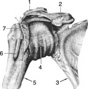
Fig. 31. Right shoulder joint (anterior view):
1 — acromion; 2 — coracoid process; 3 — scapula; 4 — articular capsule; 5 — humerus; 6 — tendon of the long head of the biceps brachii muscle; 7 — subscapularis muscle
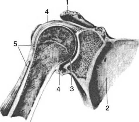
Fig. 32. Frontal section of the right shoulder joint (anterior view):
1 — acromion; 2 — scapula; 3 — joint cavity; 4 — articular capsule; 5 — tendon of the long head of the biceps brachii
The forearm (antebrachii) consists of two bones connected by a syndesmosis: the radius, located on the lateral side of the forearm, and the ulna on the medial side.
The radius (Fig. 33) is a long bone consisting of a shaft and two extremities. Its upper end features a cylindrical head with an articular facet for articulation with the capitulum of the humerus. The margin of the radial head bears an articular circumference, through which the radius articulates with the corresponding radial notch of the ulna. Below the head lies the neck, inferior to which is the radial tuberosity. The lower end of the radius is expanded and bears the carpal articular surface,
which articulates with the proximal row of carpal bones. On its medial margin is the ulnar notch for articulation with the ulnar head, while the lateral margin bears the styloid process, to which the ligaments of the hand attach. The shaft of the radius is smooth, rounded laterally and sharp medially, facing the side of articulation with the ulna.
The ulna has a triangular shaft (see Fig. 33). Its anterior and posterior surfaces, connected to the radius by a Connective Tissue membrane, form the interosseous border, located laterally relative to the radius.
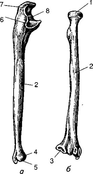
Fig. 33. Right ulna (a) and right radius (b):
1 — head of the radius; 2 — interosseous border; 3 — ulnar notch; 4 — head of the ulna; 5 — styloid process; 6 — radial notch for articulation with the radial head; 7 — olecranon process; 8 — trochlear notch; 9 — coronoid process
The upper extremity of this bone is thickened and features two processes: the posterior — the olecranon, with a rough surface for the attachment of the triceps brachii muscle, and the anterior, somewhat smaller — the coronoid process. The brachialis muscle attaches to the ulnar tuberosity located below the coronoid process. On the lateral surface of the coronoid process, There is a radial notch for articulation with the radial head. Between the olecranon and coronoid processes lies the trochlear notch, through which the ulna articulates with the trochlea of the humerus. The lower extremity of the ulna bears a head with articular surfaces — the articular circumference, by means of which it connects with the radius, as well as an inferior articular surface for articulation with the carpal bones. Posteromedially on the head lies the styloid process.
The elbow joint (articulatio cubiti) is formed by three bones: the distal end of the humerus and the proximal ends of the radius and ulna. It is a compound joint, as it incorporates three articulations: the humeroradial, humeroulnar, and proximal radioulnar joints (Fig. 34). The humeroradial joint is spheroidal in shape, allowing movement around three axes: vertical, horizontal, and sagittal. The humeroulnar joint is a uniaxial ginglymus (hinge joint), allowing movement around the frontal axis. The proximal radioulnar joint is a uniaxial pivot joint, allowing movement around the vertical axis — pronation and supination. These three joints share a common articular capsule, which attaches to the humerus in such a way that the radial and coronoid fossae are located within the joint cavity, while the olecranon fossa (posteriorly) lies partially outside the joint cavity. On the radius, the articular capsule attaches to the neck, and on the ulna, to the margins of its trochlear notch. The elbow joint is stabilized by two ligaments: the ulnar collateral and radial collateral ligaments. The annular ligament of the radius encircles the radial head and blends with the articular capsule. The elbow joint permits flexion and extension around the frontal axis, and rotation around the vertical axis, which occurs between the humerus and radius, as well as between the radius and ulna.
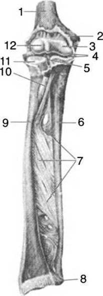
Fig. 34. Articulations of the forearm bones:
1 — humerus; 2 — articular capsule; 3 — trochlea of the humerus; 4 — cavity of the elbow joint; 5 — proximal radioulnar joint; 6 — ulna; 7 — interosseous membrane; 8 — distal radioulnar joint; 9 — radius; 10 — tendon of the biceps muscle; 11 — head of the radius; 12 — capitulum
By articulating with each other, the radius and ulna form the proximal and distal radioulnar joints of cylindrical shape, which function together as a combined pivot joint. Movements in these joints occur around a vertical axis, with the ulna remaining stationary while the radius, along with the hand, rotates around it.
The hand (manus) consists of three parts: the carpus (wrist), metacarpus, and phalanges of the digits (phalanges digitorum) (Fig. 35); it has an anterior surface — the palm (palmar) — and a posterior surface — the dorsal aspect.
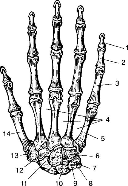
Fig. 35. Bones of the right hand (dorsal aspect):
1 — distal (nail) phalanx; 2 — middle phalanx; 3 — proximal phalanx; 4 — interosseous spaces; 5 — fifth metacarpal bone; 6 — hamate bone; 7 — capitate bone; 8 — pisiform bone; 9 — triquetrum; 10 — lunate; 11 — scaphoid; 12 — trapezoid bone; 13 — trapezium; 14 — first metacarpal bone
The carpus consists of eight bones arranged in two rows: proximal and distal. Starting from the lateral side, the proximal row includes the scaphoid, lunate, triquetrum, and pisiform bones. The first three bones are tightly bound together by ligaments. Through their articular surfaces, they form an ellipsoidal convexity, which articulates with the radius to form the radiocarpal joint. The pisiform bone articulates exclusively with the triquetrum, forming a palmar projection to which muscle tendons and ligaments attach. The distal row of carpal bones includes the trapezium, trapezoid, capitate, and hamate bones. They articulate with the proximal row of carpal bones and with the metacarpal bones.
The radiocarpal joint (articulatio radiocarpea) (Fig. 36) is ellipsoidal in shape, formed by the articular surface of the radius and the proximal row of carpal bones (excluding the pisiform). The joint is well reinforced by palmar, dorsal, and collateral ligaments. The ulna does not participate in The formation of this joint. The radiocarpal joint has two axes of rotation: sagittal and frontal. Flexion and extension are possible around the frontal axis, whereas abduction and adduction occur around the sagittal axis. Circumduction is also possible in this joint.
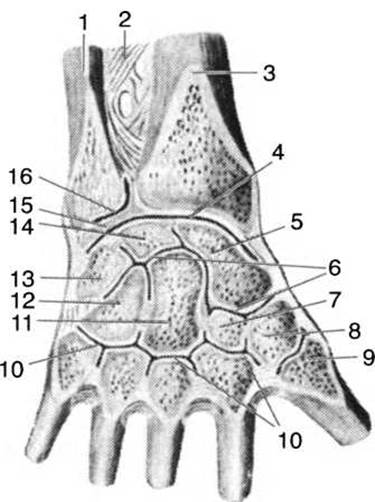
Fig. 36. Articulations of the hand (right):
1 — ulna; 2 — interosseous membrane of the forearm; 3 — radius; 4 — radiocarpal joint; 5 — scaphoid bone; 6 — intercarpal joint; 7 — trapezoid bone; 8 — trapezium bone; 9 — carpometacarpal joint of the thumb; 10 — carpometacarpal joints of digits II–V; 11 — capitate bone; 12 — hamate bone; 13 — triquetral bone; 14 — lunate bone; 15 — articular disc; 16 — distal radioulnar joint
The metacarpus consists of five long bones. Each of them has a base, shaft, and head. By means of their bases, they articulate with the carpal bones, and through their heads, with the phalanges of the fingers. The first metacarpal bone is shorter and wider than the others; it forms a saddle joint with the trapezium, providing a wide range of motion for the thumb (digit I).
Bones of the digits of the hand. Each digit (except the first) has three phalanges: proximal, middle, and distal. The thumb lacks a middle phalanx, possessing only a proximal and a distal phalanx. Each phalanx consists of a base, shaft, and head. The bases of the phalanges are directed proximally, and their heads distally. The bases and heads bear articular surfaces through which the phalanges articulate with one another and with the metacarpal bones.
The intercarpal joint (articulatio intercarpea) is formed by the two rows of carpal bones. Functionally, it is linked with the radiocarpal joint, allowing slight flexion and extension. All carpal bones are secured by short, strong ligaments.
The carpometacarpal joint (articulatio carpometacarpea) is located between the distal row of carpal bones and the bases of the II, III, IV, and V metacarpal bones. It is a slightly movable joint classified as an amphiarthrosis, reinforced by short ligaments.
The carpometacarpal joint of the thumb is formed by the articular surfaces of the Base of the I metacarpal bone and the distal surface of the trapezium. It is saddle-shaped, highly mobile, and biaxial, allowing flexion and extension, abduction and adduction of the thumb, opposition to the other digits, and circumduction.
The metacarpophalangeal joints (articulationes metacarpophalan geae) are formed by the heads of the II–V metacarpal bones and the bases of the proximal phalanges of digits I–V. Their shape is close to spherical, though the full range of a ball-and-socket joint is restricted by well-developed collateral ligaments. These joints allow movement around the frontal axis (flexion and extension) and around the sagittal axis (abduction and adduction).
The interphalangeal JOINTS OF THE hand (articulationes interphalangeae manus) are formed by the heads and bases of adjacent phalanges and are hinge-shaped, permitting movement exclusively around the frontal axis in the form of flexion and extension. The joints feature well-developed collateral stabilizing ligaments.
Last update: 08/08/2026
Editorial and Educational Adaptation: This material has been compiled based on the primary/original source text. The project team performed an editorial review, corrected technical inaccuracies, structured sections, and adapted the content for an educational format.
What was processed:
- elimination of formatting defects (OCR errors, structural breaks, corrupted characters);
- editorial organization of content;
- standardization of terminology in accordance with academic sources;
- verification of factual statements against the original source text.
All mentions of the author, publication year, and origin of the primary text have been preserved in accordance with the source.