Human Anatomy - Kotsan I. Ya. 2009
Splanchnology
Digestive system
Esophagus
The oesophagus is a direct continuation of the Pharynx. It is a cylindrical tube, flattened in the anteroposterior direction, through which food passes from the pharynx into The Stomach. In adults, the oesophagus reaches a length of 25–30 cm and a width of 3 cm. It begins at the level of the VI cervical vertebra, which corresponds to the lower edge of the laryngeal cricoid Cartilage, and ends at the level of the XI thoracic vertebra.
Since the oesophagus originates in the neck region, extends into the thoracic cavity, and then enters the Abdominal cavity through the oesophageal hiatus of the Diaphragm, topographically it is divided into three parts: cervical, thoracic, and abdominal (Fig. 145). Clinicians also distinguish an additional part between the thoracic and abdominal sections known as the diaphragmatic part.
The cervical part (pars cervicalis) of the oesophagus extends from the level of the VI cervical vertebra to the I–II thoracic vertebrae, and is located posterior and slightly to the left of the Trachea, anterior to the bodies of the aforementioned vertebrae upon the prevertebral layer of the Cervical fascia. The common carotid Arteries and recurrent laryngeal nerves run along the sides of this part of the oesophagus. The length of the cervical oesophagus is 5–8 cm.
The thoracic part (pars thoracica) of the oesophagus is the longest, passing through the entire thoracic cavity from the superior thoracic aperture to the oesophageal hiatus of the diaphragm. Its length is 15–18 cm. The thoracic oesophagus is initially located in the superior Mediastinum, then in the posterior inferior mediastinum, and is accompanied by the vagus nerves. Within the thoracic cavity, the oesophagus initially runs to the right of the aorta, then gradually shifts to its anterior surface, and prior to passing through the diaphragm, it is situated to the left of it.
The abdominal part (pars abdominalis) of the oesophagus, 1–3 cm in length, is located in the abdominal cavity beneath the diaphragm and is covered anteriorly by the left lobe of the Liver.
The lumen of the oesophagus is not uniform throughout its length.
Three anatomical constrictions are distinguished in the oesophagus — superior, middle, and inferior — along with two dilations: superior and inferior.
The superior constriction is located at the level of the VI–VII cervical vertebrae at the junction of the pharynx and oesophagus; the middle constriction — at the level of the IV–V thoracic vertebrae, where the oesophagus is crossed by the left main bronchus; the inferior constriction — at the level of the X–XI thoracic vertebrae, where the oesophagus passes through the oesophageal hiatus of the diaphragm. The superior and inferior dilations are situated between these constrictions. In addition to the three anatomical constrictions, two physiological constrictions are distinguished along the course of the oesophagus: the aortic constriction (at the crossing of the oesophagus with the aortic arch) and the cardiac constriction (at the transition of the oesophagus into the cardiac part of the stomach). Physiological constrictions exist only in living humans.
Class="center">
Fig. 145 Oesophagus (anterior view)
1 — pharynx (laryngeal part); 2 — cervical part of the oesophagus; 3 — thoracic part of the oesophagus; 4 — abdominal part of the oesophagus; 5 — cardiac orifice; 6 — diaphragm; 7 — inferior constriction of the oesophagus; 8 — middle constriction of the oesophagus
The wall of the oesophagus (approximately 4 mm thick) consists of the mucous membrane, submucosa, muscular coat, and adventitial coat (Fig. 146).
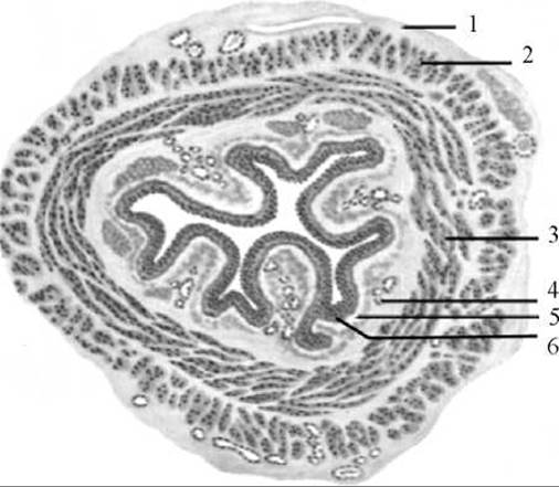
Fig. 146. Oesophagus (cross-section)
1 — outer coat (adventitia); 2 — muscular coat (longitudinal layer); 3 — muscular coat (circular layer); 4 — submucosal layer; 5 — mucous membrane; 6 — epithelium
The mucous membrane (tunica mucosa) is lined with stratified squamous epithelium. The thickness of the mucous membrane is formed by loose Fibrous Connective Tissue with a well-developed muscular lamina of the mucosa. The mucous membrane contains mucous glands, the secretion of which facilitates the passage of food through the oesophagus. In addition to mucous glands, small glands structurally similar to the cardiac Glands of the stomach are found in the abdominal and, more rarely, the thoracic PARTS OF THE oesophagus.
The submucosa (tela submucosa) is well-developed, causing the overlying mucous membrane to form longitudinal folds. Longitudinal folding is a functional adaptation of the oesophagus that facilitates the movement of fluids through the channels between the folds and the expansion of its lumen during the passage of solid food boluses. In cross-section, the lumen of the oesophagus has the shape of a stellate cleft. The submucosa houses Blood Vessels, nerve plexuses, solitary lymphatic follicles, and mucous glands, the ducts of which open onto The surface of the mucous membrane.
The muscular coat (tunica muscularis) of the oesophagus consists of two layers: an inner circular layer and an outer longitudinal layer. Vascular networks and nerve plexuses are located in the connective tissue intermuscular layer. In the upper third of the oesophagus, the muscular layers are formed by Striated Muscle tissue, which transitions into smooth muscle in the middle part, and consists exclusively of smooth musculature in the lower third.
The adventitial (outer) coat (tunica adventitia) covers the cervical and thoracic parts of the oesophagus, whereas the abdominal part of the organ is covered by the visceral Peritoneum. The adventitial coat also contains blood and Lymphatic vessels and nerve trunks that form plexuses. The adventitial coat is formed of loose fibrous connective tissue containing a small amount of elastic fibers. Via this coat, the oesophagus is anchored to adjacent surrounding Organs in the mediastinum.
The Blood supply to the oesophagus comes from several sources. Its cervical part is supplied by the inferior thyroid arteries, the thoracic part by Branches of the Thoracic Aorta, and the abdominal part by branches of the left gastric artery. Venous blood drains from the cervical region into the subclavian vein, from the thoracic region into the azygos and hemiazygos Veins, and from the abdominal region into the left gastric vein.
The lymphatic Vessels of the cervical part of the oesophagus drain into the deep lateral (jugular) Lymph Nodes, those of the thoracic part into the prevertebral and posterior mediastinal lymph nodes, and those of the abdominal part into the left gastric lymph nodes.
Sensory and parasympathetic Innervation of the oesophagus is supplied by the oesophageal branches of the Vagus nerve, while sympathetic innervation is derived from the Sympathetic trunk.
Last update: 08/08/2026
Editorial and Educational Adaptation: This material has been compiled based on the primary/original source text. The project team performed an editorial review, corrected technical inaccuracies, structured sections, and adapted the content for an educational format.
What was processed:
- elimination of formatting defects (OCR errors, structural breaks, corrupted characters);
- editorial organization of content;
- standardization of terminology in accordance with academic sources;
- verification of factual statements against the original source text.
All mentions of the author, publication year, and origin of the primary text have been preserved in accordance with the source.