Human Anatomy, Part 2 - K. A. Dyubenko, A. K. Kolomiytsev, Yu. B. Chaykovsky 2008
Special Section
Cardiovascular System, systema cardiovasculare - Special Section
Blood vessels of the systemic circulation - Arteries of the lower limb - Femoral artery
The femoral artery, a. femoralis (Figs. 51, 52, 52A), is the direct continuation of the external iliac artery. Together with the femoral vein, it passes through the lacuna vasorum onto the anteromedial surface of the thigh, where it lies within the iliopectineal sulcus, sulcus iliopectineus. Further distally, the artery courses obliquely from front to back through the femoral triangle, trigonum femorale s. Scarpi, and through the adductor canal, canalis adductorius (Hunten), reaching the popliteal fossa, where it becomes the popliteal artery, a. poplitea. The PROJECTION OF THE femoral artery onto the Skin of the thigh extends from the midpoint of the inguinal ligament to the medial epicondyle of the Femur (Keen's line).
Branches of the femoral artery
1. Superficial epigastric artery, a. epigastrica superficialis;
2. Superficial circumflex iliac artery, a. circumflexa iliacum superficialis;
3. External pudendal artery, a. pudenda externa;
4. Deep artery of the thigh, a. profunda femoris;
5. Descending genicular artery, a. genus descendens.
1. The superficial epigastric artery, a. epigastrica superficialis, originates from the femoral artery below the inguinal ligament, lig. inguinale, and ascends toward the umbilicus, where it anastomoses with the superior epigastric artery (a branch of the internal thoracic artery, a. thoracica interna) and the intercostal Arteries.
2. The superficial circumflex iliac artery, a. circumflexa ilium superficialis (Fig. 51), runs parallel to the inguinal ligament, lig. inguinale, reaching the anterior superior iliac spine, where it anastomoses with the deep circumflex iliac artery, a. circumflexa iliaca profunda (a branch of the external iliac artery), and branches of the deep artery of the thigh.
3. The external pudendal arteries, aa. pudendae externae (Figs. 51, 52), comprising 2–3 branches, course medially and superiorly, passing within the subcutaneous tissue anterior to the femoral vein to supply the Scrotum (anterior scrotal branches, rr. scrotales anteriores) or, in females, the Labia Majora (anterior labial branches, rr. labiales anteriores).
4. The deep artery of the thigh, a. profunda femoris (Figs. 51, 52), is the largest branch of the femoral artery*. It originates within the femoral triangle from the posterior aspect of the femoral artery, 5–6 cm inferior to the inguinal ligament, though a lower origin at a distance of 10–11 cm is occasionally observed. Its length ranges from 6 to 8 cm. The deep artery of the thigh serves as the main collateral pathway in The Development of collateral Circulation. When ligating the a. femoralis, the ligature should be placed as close as possible to the Water/144.html">Origin of the a. profunda femoris to avoid excluding the cutaneous branches arising from the femoral artery (a. epigastrica superficialis et a. circumflexa iliaca superficialis) from the collateral circulation (V. V. Kovanov, A. A. Travin, 1983).
* Occasionally, its diameter approaches that of the a. femoralis.
Class="center">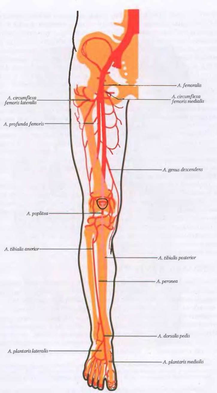
Fig. 50. Diagram of the arteries and collateral Vessels of the lower limb
The deep artery of the thigh gives rise to several branches:
- The medial femoral circumflex artery, a. circumflexa femoris medialis, originates from the posterior wall of the deep artery of the thigh*, courses medially around the femoral neck, and divides into: a superficial branch, r. superficialis; a deep branch, r. profundus; an acetabular branch, r. acetabularis; an ascending branch, r. ascendens; and a descending branch, r. descendens. The medial femoral circumflex artery anastomoses with the obturator, inferior gluteal, and lateral femoral circumflex arteries.
- The lateral femoral circumflex artery, a. circumflexa femoris lateralis, arises from the lateral aspect of the deep artery of the thigh. Passing deep to the rectus femoris Muscle, m. rectus femoris, it divides into: an ascending branch, r. ascendens, which winds around the femoral neck to anastomose with the medial circumflex artery; a transverse branch, r. transversus, which supplies the Muscles of the middle portion of the thigh; and a descending branch, r. descendens, which supplies the anterior thigh muscles.
- The perforating arteries, aa. perforantes (I–III arteries)* **, represent the terminal branches of the deep artery of the thigh. They originate from the distal portion of the a. profunda femoris, pierce the adductor muscles to reach the posterior aspect of the thigh, and supply the adductors, Posterior Thigh Muscles, skin, and the nutrient Arteries of the femur, aa. nutriendae femoris. The perforating branches anastomose with branches of the deep artery of the thigh.
- The descending genicular artery, a. descendens genicularis, originates from the terminal segment of the femoral artery within the adductor canal, canalis adductorius. Accompanied by the saphenous nerve, n. saphenus, it emerges medially above the knee joint, supplies the quadriceps femoris muscle, and participates in The formation of the arterial network of the knee joint.
Last update: 08/08/2026
Editorial and Educational Adaptation: This material has been compiled based on the primary/original source text. The project team performed an editorial review, corrected technical inaccuracies, structured sections, and adapted the content for an educational format.
What was processed:
- elimination of formatting defects (OCR errors, structural breaks, corrupted characters);
- editorial organization of content;
- standardization of terminology in accordance with academic sources;
- verification of factual statements against the original source text.
All mentions of the author, publication year, and origin of the primary text have been preserved in accordance with the source.