Human Anatomy, Part 1 - K. A. Dyubenko, A. K. Kolomiytsev, Yu. B. Chaykovsky 2002
Special Part
Myology, myologia [the study of muscles] — Muscles and Fascia of the Trunk
Muscles and Fascia of the Thorax — Diaphragm
The Diaphragm (diaphragm, thoraco-abdominale) is an unpaired Muscle that forms a thin plate (tendinous in the center and muscular at the periphery) inserted into the inferior thoracic aperture (Fig. 131).
Class="center">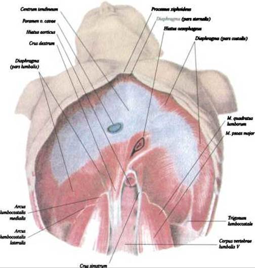
Fig. 131. The diaphragm (diaphragma), inferior view (after V. P. Vorobyov)
The diaphragm is dome-shaped, bulging superiorly into the thoracic cavity and separating it from the Abdominal cavity. The muscle bundles originate from the bony and cartilaginous PARTS OF THE inferior thoracic aperture and from the lumbar vertebrae. The bundles converge medially and terminate in the central tendon (centrum tendineum). The diaphragm is divided into three parts:
1. The lumbar part (pars lumbalis diaphragmatis).
2. The sternal part (pars sternalis diaphragmatis).
3. The costal part (pars costalis diaphragmatis).
1. The lumbar part (pars lumbalis diaphragmatis), the posterior region of the diaphragm, occupies the area of the upper four lumbar vertebrae and is divided into two crura: the right crus (crus dextrum) and the left crus (crus sinistrum). The crura originate from the anterolateral surface of the bodies of lumbar vertebrae I–III (I–IV on the right) and from the medial and lateral arcuate ligaments (lig. arcuatum mediale et laterale). The medial arcuate ligament (lig. arcuatum mediale) spans across the anterior surface of the psoas major muscle from the body to the transverse process of the first lumbar vertebra. The lateral arcuate ligament (lig. arcuatum laterale) arches over the quadratus lumborum muscle from the transverse process of the first lumbar vertebra to the XII rib. The medial muscle bundles of the diaphragmatic crura course superiorly, converge, and form the aortic hiatus (hiatus aorticus), through which the aorta and the Thoracic duct (ductus thoracicus) pass. Slightly superiorly, the medial muscle bundles form the esophageal hiatus (hiatus oesophageus), through which the Esophagus and vagus nerves pass. The so-called sliding (hiatal) hernias pass through the esophageal hiatus, occurring in 40% of the population during X-ray examinations (Jon A. Reid, 1997).
In addition, the crura of the lumbar part contain two paired clefts: a cleft through which the azygos vein (v. azygos) and the greater and lesser splanchnic nerves (nn. splanchnicus major et minor) pass on the right, and the hemiazygos vein (v. hemiazygos) and the greater and lesser splanchnic nerves (nn. splanchnicus major et minor) on the left, as well as a cleft transmitting the Sympathetic trunk (truncus sympaticus).
2. The sternal part (pars sternalis diaphragmatis) consists of small bundles originating from the xiphoid process. Between the sternal part and the costal part (pars costalis diaphragmatis), as well as between the costal part and the lumbar part (pars lumbalis diaphragmatis), there are paired, more or less distinct triangular clefts: the sternocostal triangles and lumbocostal triangles (trigonum sternocostales et trigonum lumbocostales). In these areas, the thoracic and abdominal cavities are separated only by the serous membrane. Occasionally, these triangles serve as sites for The formation of diaphragmatic hernias (congenital or acquired).
The central tendon (centrum tendineum) is a dense, shiny plate formed by the interlacing of fibrous bundles that are continuations of the muscle fibers. In the posterior region to the right of the midline lies the opening for the INFERIOR VENA CAVA (foramen venae cavae inferioris), which is bounded on all sides by fibrous tissue and transmits the inferior vena cava (v. cava inferior).
The thoracic and abdominal surfaces of the diaphragm are covered with fasciae: superiorly by the endothoracic fascia (fascia endothoracica) and inferiorly by the endoabdominal fascia (fascia endoabdominalis), which in turn are covered by serous membranes. The following Organs lie adjacent to the diaphragm: superiorly, in the region of the central tendon, The Heart; laterally, the Lungs; and inferiorly, the Liver, Stomach, Adrenal Glands, Kidneys, and Spleen. Two domes can be distinguished in the diaphragm: the right and the left. The apices of the domes reach the level of the fourth intercostal space on the right, and the fifth intercostal space on the left.
3. The costal part (pars costalis diaphragmatis) is a broad section of the diaphragm that originates by means of digitations from the lower six Ribs. These muscle bundles arch and terminate at the anterior margins of the central tendon (centrum tendineum).
Function: The diaphragm is the primary respiratory muscle that changes the volume of the thoracic cavity. Upon contraction, it descends by 1–3 cm, flattening its shape and facilitating inspiration. The diaphragm drives the abdominal type of Respiration. The contraction rate of the diaphragm in adults ranges from 16 to 20 breaths per minute. In addition to its respiratory function, the diaphragm facilitates venous return to The Heart and the emptying of hollow organs.
Blood supply: Pericardiacophrenic, superior phrenic, inferior phrenic, musculophrenic, and posterior intercostal Arteries (aa. pericardiacophrenicae, phrenicae superiores, phrenicae inferiores, musculophrenicae, intercostales posteriores).
Innervation: Phrenic nerves (nn. phrenici, C III–C VI).
Last update: 08/08/2026
Editorial and Educational Adaptation: This material has been compiled based on the primary/original source text. The project team performed an editorial review, corrected technical inaccuracies, structured sections, and adapted the content for an educational format.
What was processed:
- elimination of formatting defects (OCR errors, structural breaks, corrupted characters);
- editorial organization of content;
- standardization of terminology in accordance with academic sources;
- verification of factual statements against the original source text.
All mentions of the author, publication year, and origin of the primary text have been preserved in accordance with the source.