MEDICAL BIOLOGY, HUMAN ANATOMY, PHYSIOLOGY AND PATHOLOGY - Y.I. Fedonyuk 2010
ANATOMY, PHYSIOLOGY, PATHOLOGY
SECTION 5. BLOOD AND LYMPH CIRCULATION PROCESSES AND THEIR PATHOLOGY
VESSELS OF THE SYSTEMIC CIRCULATION
2. VEINS OF THE SYSTEMIC CIRCULATION
2.4. CAVOCAVAL AND PORTOCAVAL ANASTOMOSES
Tributaries of the portal vein anastomose with tributaries of the Veins belonging to the systems of the superior and inferior venae cavae, forming the so-called portocaval anastomoses (Fig. 5.37), which are of great clinical significance.
Class="center">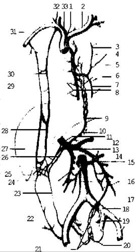
Fig. 5.37. Diagram of anastomoses between the portal, superior, and inferior venae cavae
Portocaval anastomoses are located in the abdominal region of the Esophagus between the roots of the left gastric vein, which empties into the portal vein, and the esophageal VEINS OF THE SUPERIOR VENA CAVA system.
In the lower part of the rectum, there are anastomoses between the superior rectal vein—which drains through the superior mesenteric vein into the portal vein—and the middle rectal vein, a branch of the internal iliac artery of the INFERIOR VENA CAVA system.
When Blood flow through the portal Venous system is obstructed (e.g., due to Liver cirrhosis or compression of the portal vein by a tumor), blood is diverted via these anastomoses from the esophagus and rectum into the superior and inferior venae cavae systems. This results in the dilation and engorgement of these anastomotic channels, frequently precipitating severe hemorrhages from the cardiac region of the esophagus and the rectum.
Within the thickness of the anterior abdominal wall, around the umbilicus, paraumbilical veins (tributaries of the portal vein) anastomose with the superior epigastric vein of the inferior vena cava system, thus forming a portocaval anastomosis.
Under conditions of impaired blood flow through the portal system, these anastomoses dilate, making the venous pattern visible through the Skin. This network of dilated, tortuous veins in the umbilical region is known as "caput medusae".
In the region of the posterior abdominal wall, There is a cavocaval anastomosis between the lumbar veins of the inferior vena cava system and the ascending lumbar veins, which give rise to the azygos and hemiazygos veins.
Last update: 08/08/2026
Editorial and Educational Adaptation: This material has been compiled based on the primary/original source text. The project team performed an editorial review, corrected technical inaccuracies, structured sections, and adapted the content for an educational format.
What was processed:
- elimination of formatting defects (OCR errors, structural breaks, corrupted characters);
- editorial organization of content;
- standardization of terminology in accordance with academic sources;
- verification of factual statements against the original source text.
All mentions of the author, publication year, and origin of the primary text have been preserved in accordance with the source.