ATLAS OF HUMAN ANATOMY - G.L. Bilich - 2014
APPARATUS OF LOCOMOTION
SKELETAL SYSTEM
BONES OF THE UPPER LIMB
Bones of the Free Upper Limb
The humerus (humerus, s. os brachii) is a long tubular bone consisting of a shaft (corpus humeri) and two extremities: a thickened upper (proximal) end and an expanded lower (distal) end (Fig. 88). The upper extremity bears the rounded HEAD of the humerus (caput humeri), which articulates with the glenoid cavity of the scapula to form the shoulder joint (Fig. 89). Below the head lies the anatomical neck (collum anatomicum). Laterally adjacent to the head are the greater tubercle (tuberculum majus), to which the supraspinatus, infraspinatus, and teres minor Muscles attach, and the lesser tubercle (tuberculum minus), situated anterior to the greater tubercle. The subscapularis Muscle attaches to the lesser tubercle. Inferior to the greater tubercle runs its crest (crista tuberculi majoris), which serves as the insertion site for the pectoralis major muscle. Extending downward from the lesser tubercle is the crest of the lesser tubercle (crista tuberculi minoris), to which the tendon of the latissimus dorsi and the teres major muscle attach. Between the tubercles and inferior to them, nestled between their crests, lies the intertubercular sulcus (sulcus intertubercularis), which lodges the tendon of the long head of the biceps brachii. Lateral to the crest of the greater tubercle is the deltoid tuberosity (tuberositas deltoidea), where the deltoid muscle attaches. The narrowest region between the humeral head and its shaft is termed the surgical neck (collum chirurgicum), which is the most common site for bone fractures.
Class="center">
Fig. 87. Right scapula (A — posterior view, B — anterior view):
1 — Inferior angle; 2 — Medial border; 3 — Superior angle; 4 — Supraspinous fossa; 5 — Superior border; 6 — Suprascapular notch; 7 — Spine of scapula; 8 — Coracoid process; 9 — Acromion; 10— Acromial angle; 11 — Glenoid cavity; 12 — Infraglenoid tubercle; 13 — lnfraspinous fossa; 14 — Lateral border; 15 — Neck of scapula; 16— Lateral angle; 17—Supraglenoid tubercle; 18— Subscapular fossa

Fig. 88. Right humerus (A — posterior view, B — anterior view, C, D, E — INJURIES OF THE upper right humeral epiphysis, anterior view):
1 — Trochlea; 2 — Medial epicondyle; 3 — Groove for ulnar nerve; 4 — Media! supraepicondylar ridge; Medial supracondylar ridge; 5 — Olecranon fossa; 6 — Medial border; 7 — Shaft of humerus; Body of humerus, posterior surface; 8 — Surgical neck; 9 — Anatomical neck; 10 — Head of humerus; 11 — Greatcr tubercle; 12 — Radial groove; Groove for radial nerve: 13 — Lateral margin; 14 — Radia! fossa; 15 — Medial supraepicondylar ridge; Medial supracondylar ridge; 16 — Capitulum; 17 — Lateral epicondyle; 18 — Deltoid tubcrosity; 19 —Crest ofgreater tubercle; Lateral lip; 20 — Intertubercular sulcus; Bicipital groove; 21 — Lesser tubercle: 22 — Crest of lesser tubercle; Medial lip; 23 — Anteromcdial surface; 24 — Anterolateral surface; 25 — Coronoid fossa
The upper section of the humeral shaft is cylindrical, whereas the lower section is triangular in cross-section. In this region, the bone presents three surfaces: posterior (facies posterior), medial anterior (facies medialis anterior), and lateral anterior (facies lateralis anterior). Approximately at the middle of the humerus runs the radial groove (sulcus nervi radialis), which begins on the medial side of the bone, spirals posteriorly around the shaft, and terminates inferiorly at its lateral border.
The lower epiphysis of the humerus is expanded, flattened, slightly anteriorly curved, and forms the humeral condyle (condylus humeri) (Fig. 90). The medial part of the condyle—the trochlea of the humerus (trochlea humeri)—articulates with the trochlear notch of the ulna. The lateral part of the condyle forms the capitulum of the humerus (capitulum humeri), which articulates with the articular fovea of the head of the radius. Anteriorly, just above the humeral trochlea, lies the coronoid fossa (fossa coronoidea), which accommodates the coronoid process of the ulna during flexion at the elbow joint. Above the capitulum, the radial fossa (fossa radialis) is visible. Posteriorly, superior to the condyle, is the olecranon fossa (fossa olecrani), which receives the olecranon process of the ulna during extension of the forearm (straightening of the upper limb). Between the olecranon fossa and the coronoid fossa lies a thin bony partition that occasionally features an aperture.
Elevations known as epicondyles are located on the medial and lateral sides above the humeral condyle. The ulnar nerve groove (sulcus nervi ulnaris) runs along the posterior surface of the medial epicondyle (epicondylus medialis). Superiorly, this epicondyle transitions into the medial supracondylar ridge (crista supracondylaris medialis), which forms the medial border of the humerus. The smaller lateral epicondyle (epicondylus lateralis) continues superiorly into the lateral supracondylar ridge (crista supracondylaris lateralis), forming the lateral margin of the bone.

Fig. 89. Head of the right humerus:
1 — Lesser tubercle; 2 — Intertubercular sulcus; Bicipital groove; 3 — Greater tubercle; 4 — Anatomical neck; 5 — Head of humerus
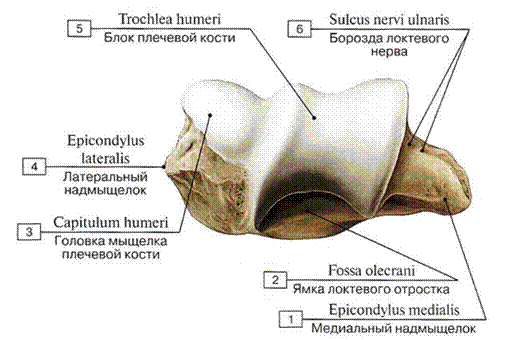
Fig. 90. Condyle of the right humerus:
1 — Medial epicondyle; 2 — Olecranon fossa; 3 — Capitulum; 4— Lateral epicondyle; 5 — Trochlea; 6 — Groove for ulnar nerve

Fig. 91. Radiograph of the humerus, mediolateral projection:
1 — clavicle; 2 — coracoid process; 3 — acromion of the scapula; 4 — glenoid cavity of the scapula; 5 — head of the humerus; 6 — surgical neck of the humerus; 7 — diaphysis of the humerus; 8 — coronoid fossa of the humerus; 9 — superimposed image of the capitulum and trochlea of the humerus; 10 — olecranon fossa of the humerus; 11 — radius; 12 — ulna (after A.Yu. Vasilyev)
An anatomical variation is the presence of an aperture above the medial epicondyle of the humerus, known as Knox's foramen (syn.: supracondylar process foramen, foramen supracondylare humeri). In some cases, a hook-like downward-curved supracondylar process (processus supracondylaris) of varying length is observed. Occasionally, its tip is connected to the medial epicondyle by a ligament that may undergo ossification, thereby forming a supracondylar foramen—a feature common to many mammals (Robert Knox, 1791–1862, British physician).
An anatomical landmark used in the radiographic evaluation of patients with recurrent shoulder dislocation is Adams' groove, a longitudinal defect on the posterolateral surface of the humeral head (named after William Adams, 1760–1829, an English surgeon).
Fig. 91 shows radiographs of the humerus.
The forearm (antebrachium) comprises the ulna, located medially, and the radius, located laterally (Figs. 92, 93). Both bones contact each other only at their extremities. Between the shafts of these bones lies the interosseous space of the forearm (spatium interosseum antebrachii). Each bone has a shaft and two epiphyses: proximal and distal. The sharp interosseous border (margo interosseus) of both the ulna and the radius faces the interosseous space, providing attachment for the fibrous interosseous membrane of the forearm.
The ulna (see Fig. 92) features a thickened upper (proximal) end bearing the trochlear notch (incisura trochlearis) for articulation with the trochlea of the humerus, thereby forming the humeroulnar joint. The posterior margin of the notch is bounded by the stout olecranon. Anterior to the notch lies the small coronoid process (processus coronoideus). On the lateral side of the Base of the coronoid process, the radial notch (incisura radialis) is visible, which articulates with the head of the radius (forming the humeroradial joint). Distal to the coronoid process is the ulnar tuberosity (tuberositas ulnae), to which the brachialis muscle attaches. The slender lower (distal) end of the ulna terminates in the ulnar head (caput ulnae), which features an articular circumference (circumferentia articularis) on its lateral surface for articulation with the radius, thus forming the distal radioulnar joint. The ulnar styloid process projects from the medial side of the head.

Fig. 92. Right ulna (A — anterior view, B — lateral view, C — posterior view):
1 — Trochlear notch; 2 — Coronoid process; 3 — Radial notch; 4 — Tuberosity of ulna; 5 — Shaft of ulna; Body of ulna, anterior surface; 6 — Interosseous border; 7 — Articular circumference; 8 — Head of ulna; 9 — Ulnarstyloid process; 10 — Olecranon; 11 — Posterior border; 12— Shaft of ulna; Body of ulna, posterior surface; 13 — Shaft of ulna; Body of ulna, medial surface
At its proximal end, the radius features the radial head (caput radii) with a depressed articular fovea (fovea articularis) for articulation with the capitulum of the humerus. Below the head lies the neck (collum radii). The lateral surface of the head bears the articular circumference (circumferentia articularis), which forms a joint with the radial notch of the ulna (the proximal radioulnar joint). Immediately distal to the neck on the anteromedial surface of the bone is the radial tuberosity (tuberositas radii), where the tendon of the biceps brachii inserts. The expanded distal end of the radius presents the ulnar notch (incisura ulnaris) on its medial side for articulation with the head of the ulna, while its posterior surface features longitudinal grooves for the tendons of the extensor Muscles of the wrist and fingers (see Fig. 93).

Fig. 93. Right radius (A — anterior view, B — medial view, C — posterior view):
1 — Head of radius, articular eircumference; 2 — Articular fovea; 3 — Neck of radius; 4 — Radial tuberosity; 5 — Anterior border; 6 — Inlerosseous border; 7 — Shaft of radius; Body of radius, anterior surface; 8 — Carpal articular surface; 9 — Radial styloid process; 10 — Shaft of radius; Body of radius, posterior surface; 11 — Ulnar notch; 12 — Posterior border; 13 — Shaft of radius; Body of radius, lateral surface; 14 — Dorsal tubcrcle

Fig. 94. Radiograph of the BONES OF THE forearm, anteroposterior view:
1 — radiocarpal joint; 2 — radial styloid process; 3 — ulnar styloid process; 4 — distal radioulnar joint; 5 — radial shaft; 6 — ulnar shaft; 7 — radial tuberosity; 8 — neck of radius; 9 — coronoid process of ulna; 10 — head of radius; 11 — olecranon; 12 — humerus (according to A. Yu. Vasilyev)
On the distal surface of the distal end of the radius lies the concave carpal articular surface (facies articularis carpea), which articulates with the bones of the proximal carpal row (the scaphoid and lunate bones) to form the radiocarpal joint. The styloid process (processus styloideus) projects from the lateral side of the distal end of the radius. On the posterior surface of the lower end of the radius, There is a bony prominence bounding the groove for the tendon of the extensor pollicis longus—Lister's tubercle (dorsal tubercle) (named after Joseph Lister, 1827–1912, an English surgeon and anatomist).
Fig. 94 shows a radiograph of the forearm bones.
The Skeleton of the hand (manus) comprises the Carpal Bones (ossa carpi), Metacarpal bones (ossa metacarpi), and the bones of the digits—the Phalanges of the hand (phalanges digitorum manus) (Figs. 95, 96).
The carpus is formed by eight short (spongy) bones arranged in two rows. In the proximal row, from the radial to the ulnar side, lie the scaphoid, lunate, triquetrum, and pisiform bones. In the distal row, in the same direction, lie the trapezium (greater multangular), trapezoid (lesser multangular), capitate, and hamate bones. Each of these bones possesses articular surfaces for articulation with adjacent bones (Fig. 97).
The scaphoid bone (os scaphoideum), curved proximally, features a tubercle directed toward the palm. The lunate bone (os lunatum) is also curved proximally and concave on its distal surface. The triquetrum (os triquetrum) bears an articular surface on its anterior aspect for articulation with the pisiform bone. The pisiform bone (os pisiforme), of irregular rounded shape, is the smallest carpal bone and is embedded within the tendon of the flexor carpi ulnaris (acting as a sesamoid bone). The proximal surfaces of the scaphoid, lunate, and triquetrum participate in The formation of the radiocarpal joint, whereas their distal surfaces face the bones of the second carpal row.
The trapezium (os trapezium), cuboidal in shape, features a saddle-shaped articular surface for articulation with the base of the first metacarpal bone ($I$). The palmar surface of the trapezium presents a groove that lodges the tendon of the flexor carpi radialis. The trapezoid bone (os trapezoideum) resembles the trapezium in shape, although it is smaller. The capitate bone (os capitatum) is the largest of the carpal bones; its rounded head is directed proximally and slightly laterally. The hamate bone (os hamatum) features a hook (hamulus ossis hamati) near the ulnar margin of its palmar surface, curved toward the radial side.

Fig. 95. Bones of the right hand, palmar surface:
1 — Ulna; 2 — Head of ulna; 3 — Ulnar styloid process; 4 — Lunate; 5 — Triquetrum; 6 — Pisiform; 7 — Hamate; 8 — Hook of hamate; 9 — Carpalbones; 10— Base of metacarpal; 11— Shaftofmetacarpal; Body of metacarpal; 12— Headofmetacarpal; 13— Metaearpals [I—V]; 14 — Base of phalanx; 15 — Shaft of phalanx: Body of phalanx; 16 — Head of phalanx; 17— Phalanges; 18 — Tuberosity of distal phalanx; 19 — Distal phalanx [II]; 20 - Middle phalanx [ІІ]; 21 — Proximal phalanx [II]; 22 — Distal phalanx [I]; 23 — Proximal phalanx [I]; 24 —Scsamoid bones; 25— Metacarpal [1]; 26 — Trapezoid; 27—Trapezium; 28— Trapezium, tubercle; 29 — Tubercle ofscaphoid; 30— Capitate; 31 — Scaphoid; 32 — Radius

Fig. 96. Bones of the right hand, dorsal surface:
1 — Radius; 2 — Lunate; 3 — Radial styloid process; 4 — Scaphoid; 5 — Trapezium; 6 — Carpometacarpal joint [I]; 7 — Trapezoid; 8 — Interphalangeal joints of hand (proximal); 9 — Interphalangeal joints of hand (distal); 10 — Metacarpophalangeal joints; 11 — Capitate; 12 — Hamate; 13 — Triquetrum; 14 — Ulnar styloid process; 15 — Ulna

Fig. 97. Bones of the metacarpus and carpus, right hand (A — distal epiphyses of the forearm bones and carpal bones, B — view of the hand after removal of the forearm bones):
1 — Radius; 2 — Dorsal tubercle; 3 — Radial styloid process; 4 — Trapezium; 5 — Trapezoid; 6 — Metacarpals [I—V]; 7 — Capitate; 8 — Hamate; 9 — Triquetrum; 10 — Lunate; 11 — Ulnar styloid process; 12 — Scaphoid; 13 — Ulna; 14 — Carpal groove; 15 — Trapezium, tubercle; 16 — Pisiform
The carpal bones, arranged in two rows, form a bony arch whose convex aspect faces dorsally and whose concave aspect faces palmarly (anteriorly). As a result, the carpal groove (sulcus carpi) is formed on the palmar surface, bounded radially by the tubercles of the scaphoid and trapezium, and ulnarly by the hook of the hamate and the pisiform bone.
The metacarpus comprises five (I—V) short tubular metacarpal bones (ossa metacarpalia) of triangular shape (see Fig. 97). Each metacarpal bone consists of a base (basis), a body (corpus), and a head (caput). The epiphyses of each metacarpal bone are thickened, so that interosseous spaces remain between the bodies when the bones articulate with one another. On the palmar aspect, the bodies of the metacarpals are slightly concave, whereas on the dorsal aspect they are slightly convex. The bases of the II—V metacarpals feature flat articular surfaces on their proximal ends for articulation with the bones of the distal carpal row. The first metacarpal bone (os metacarpale I) is shorter and thicker than the others. Its base bears a saddle-shaped articular surface for articulation with the trapezium, forming the carpometacarpal joint of the thumb. The second metacarpal bone (os metacarpale II) is the longest; its articular surface is divided into two parts for articulation with the trapezium and trapezoid bones. The dimensions of the remaining metacarpals gradually decrease toward the fifth metacarpal. The bases of the II to V metacarpals possess lateral articular surfaces designed for articulation with one another. The hemisphere-shaped heads of the metacarpals form convex articular surfaces for connection with the proximal phalanges of the digits.
Near the base of the V metacarpal bone, an accessory carpal bone often develops — the Vesalian bone (Andreas Vesalius, 1515–1564, a Renaissance scholar and founder of modern anatomy).
Bones of the digits (phalanges). The thumb (pollex, s. digitus primus) is the shortest and thickest; it is followed by II — the index finger (index, s. digitus secundus); III — the middle finger, the longest (digitus medius, s. tertius); IV — the ring finger (digitus anularis, s. quartus); and V — the little finger (digitus minimus, s. quintus). The phalanges of the fingers (phalanges digitorum) are short tubular bones. Each digit, except the first (thumb), has three phalanges: the proximal phalanx (phalanx proximalis), which is the longest; the middle phalanx (phalanx media); and the shortest, the distal phalanx (phalanx distalis). The thumb, being the thickest, has only two phalanges — proximal and distal.
One distinguishes the base of the phalanx (basis phalangis), the body of the phalanx (corpus phalangis), and the head of the phalanx (caput phalangis). The bodies of the proximal and middle phalanges are convex dorsally and slightly concave palmarly. The base of each proximal phalanx features an articular facet for articulation with the corresponding metacarpal bone. The bases of the middle and distal phalanges are provided with pulley-shaped (trochlear) articular surfaces for articulation with the heads of the proximally positioned phalanges. The extremities of each nail (distal) phalanx are flattened and bear the tuberosity of the distal phalanx (tuberositas phalangis distalis).
Fig. 98 shows a radiograph of the hand.

Fig. 98. Radiograph of the hand, palmar projection:
1 — distal phalanx; 2 — distal interphalangeal joint; 3 — proximal interphalangeal joint; 4 — middle phalanx; 5 — proximal phalanx; 6 — head of proximal phalanx; 7 — base of proximal phalanx; 8 — metacarpophalangeal joint; 9 — metacarpal bone; 10 — head of metacarpal bone; 11 — base of metacarpal bone; 12 — sesamoid bone; 13 — trapezium; 14 — trapezoid; 15 — capitate; 16 — hamate; 17 — hook of hamate; 18 — scaphoid; 19 — lunate; 20 — triquetrum; 21 — pisiform; 22 — radial styloid process; 23 — ulnar styloid process; 24 — distal radioulnar joint (according to A. Yu. Vasilyev)
Last update: 08/08/2026
Editorial and Educational Adaptation: This material has been compiled based on the primary/original source text. The project team performed an editorial review, corrected technical inaccuracies, structured sections, and adapted the content for an educational format.
What was processed:
- elimination of formatting defects (OCR errors, structural breaks, corrupted characters);
- editorial organization of content;
- standardization of terminology in accordance with academic sources;
- verification of factual statements against the original source text.
All mentions of the author, publication year, and origin of the primary text have been preserved in accordance with the source.