BIOLOGY Volume 3 - A Guide to General Biology - 2004
23. THE CONTINUITY OF LIFE
23.6. DNA
23.6.2. DNA Replication
The double-helix model of Introduction/20.html">DNA Structure proposed by Watson and Crick is described in Section 3.6.3. One of the most appealing features of this model is that it simultaneously suggests how METABOLISM/36.html">DNA Replication might occur. Watson and Crick hypothesized that the two strands forming the helix could unwind and separate, serving as templates to which a complementary chain of NUCLEOTIDES is assembled via base pairing. Thus, two identical copies are generated from each original DNA molecule.
In 1956, Kornberg successfully demonstrated the in vitro synthesis of a DNA molecule using a single DNA strand as a template. Kornberg isolated and purified an enzyme from E. coli capable of linking free nucleotides together in the presence of ATP as an energy source, forming a complementary DNA strand. He named this enzyme DNA polymerase. As subsequent experiments showed, the nucleotides utilized within The Cell carry two additional phosphate groups, which serves to activate them. As each nucleotide is attached to the growing DNA chain, the two extra phosphate groups are cleaved off. The energy released in this process is used by the remaining phosphate group of the nucleotide to form a bond with the sugar residue of the adjacent nucleotide. The replication process is illustrated in Fig. 23.20. It begins with the unwinding of the DNA double helix, a process controlled by the enzyme helicase. Next, DNA polymerase binds to the single-stranded DNA and begins moving along the chain. Whenever it reaches the next base in the DNA chain, free nucleotides approach the chain, and the one containing the complementary base forms Hydrogen Bonds with it. The free nucleotide is held in place by the enzyme until it joins the preceding nucleotide, thereby extending the new DNA strand. This extension can occur exclusively in the 5' → 3' direction. As can be seen in Fig. 23.20, this means that only One DNA strand (the top one in the figure) can be copied continuously, since the DNA polymerase moves in the same direction as the unwinding enzyme. This process is referred to as continuous replication. Copying of the other strand (the bottom one in Fig. 23.20) must be initiated repeatedly because the DNA polymerase must move away from the unwinding enzyme in the 5' → 3' direction. As a result, small gaps appear in the strand, as DNA polymerase cannot link the 3' end of one newly synthesized DNA segment to the 5' end of the next. To bridge this gap, another enzyme is required—DNA ligase. This type of replication is called discontinuous.
Class="center">
Fig. 23.20. DNA replication. A simplified diagram showing the main principles. Other molecules and Enzymes not shown in the diagram also participate in replication.
Evidence for Semiconservative Replication
The mode of DNA replication proposed by Watson and Crick and shown in Fig. 23.20 is known as semiconservative replication, because each new double helix retains one of the two strands from the original parental DNA double helix. This mechanism is based on data obtained by Meselson and Stahl in a series of classic experiments in 1958. E. coli Cells contain a single circular chromosome; by culturing these cells for many generations in a medium containing a heavy isotope of nitrogen (15N), all of their DNA became labeled with this isotope. The cells with labeled DNA were then transferred to a medium containing the common nitrogen isotope 14N. After a period corresponding to the generation time of E. coli (50 min at 36 °C)—the time required for one Cell Division and one round of DNA replication—cell samples were collected, their DNA was extracted, and it was centrifuged for 20 h at 40,000 g in a cesium chloride (CsCl) solution. During centrifugation, the heavy CsCl molecules began to sediment, establishing a density gradient that increased from the top to the bottom of the tube. The DNA banded at a position where the density of the CsCl solution matched its own density. When examined under ultraviolet light, the DNA in the centrifuge tube appeared as a narrow band. The positions of the DNA bands extracted from cells cultured in 15N and 14N media, along with the interpretation of the results, are presented in Fig. 23.21. A schematic of semiconservative replication is included in Fig. 23.22. These experiments provided compelling evidence that DNA replication proceeds via a semiconservative mechanism.
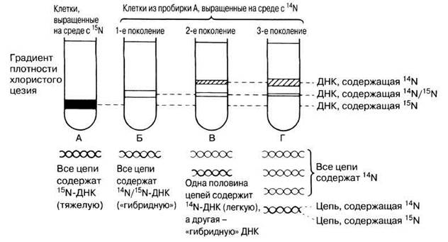
Fig. 23.21. Results of the Meselson and Stahl experiments and their interpretation. The widths of the DNA bands in the centrifuge tubes reflect the relative quantities of Different types of DNA molecules. In tube B, the band width ratio is 1:1, whereas in tube D, it is 3:1.
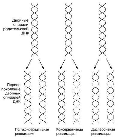
Fig. 23.22. Diagram illustrating the three theories of DNA replication.
23.2. Three hypotheses were put forward to explain The process of DNA replication. One of these is known as semiconservative replication and is described above. The other two are known as conservative replication and dispersive replication.
All three hypotheses are illustrated in Fig. 23.22.
Draw diagrams showing the distribution of different types of DNA in a density gradient that Meselson and Stahl would have observed for the first two generations of cells if the conservative and dispersive replication hypotheses were correct.
Last update: 06/08/2026
Editorial and Educational Adaptation: This material has been compiled based on the primary/original source text. The project team performed an editorial review, corrected technical inaccuracies, structured sections, and adapted the content for an educational format.
What was processed:
- elimination of formatting defects (OCR errors, structural breaks, corrupted characters);
- editorial organization of content;
- standardization of terminology in accordance with academic sources;
- verification of factual statements against the original source text.
All mentions of the author, publication year, and origin of the primary text have been preserved in accordance with the source.