BIOLOGY Volume 2 - A Guide to General Biology - 2004
13. TRANSPORT IN PLANTS
13.1. Plant Water Relations
13.1.7. Osmosis and Plant Cells
Fig. 13.1 illustrates semi-permeable membranes, which play a crucial role in the Water relations of a plant Cell. The Cell wall is generally fully permeable to any dissolved molecules; therefore, it cannot be considered an osmotic barrier. Plant Cells often contain a large central vacuole whose contents—The Cell sap—affect the overall osmotic potential of the system. In general, the cell's water relations depend on two membranes: the Plasmalemma, which surrounds the Cytoplasm externally, and the tonoplast, which encloses the vacuole.
Class="center">
Fig. 13.1. Semi-permeable membranes of a typical plant cell. The outer cell membrane (plasmalemma) is usually tightly pressed against the cell wall.
If a plant cell comes into contact with a solution whose water potential is lower than that of its internal contents (e.g., concentrated sugar syrup; see Experiment 13.1), water will move out of the cell via osmosis through the plasmalemma (Fig. 13.2). Water is first lost from the cytoplasm and subsequently from the vacuole, exiting through the tonoplast. The protoplast—that is, the living Contents of the plant cell enclosed by the cell wall—shrinks and pulls away from the wall, effectively contracting inside it. This process is called plasmolysis, and a cell in this state is said to be plasmolysed. The moment when the protoplast still adheres to the cell wall but has ceased to exert pressure on it is termed incipient plasmolysis. At this point, the cell loses turgor, becoming flaccid. Water continues to leave the protoplast until The water potential of its contents matches that of the surrounding solution. An equilibrium is then established, and the shrinkage of the protoplast stops.
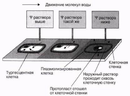
Fig. 13.2. The Effect of solutions of varying concentrations on plant cells. In a solution with a higher water potential than the cell (a hypotonic environment), water enters the cell via osmosis, causing it to swell (becoming turgid). If the solution's water potential is lower than that of the cell (a hypertonic environment), water leaves the cell via osmosis, and the living part of the cell (the protoplast) pulls away from the cell wall, following the contracting vacuole (plasmolysis). If the water potentials of the cell and the solution are equal (an isotonic environment), no changes occur.
13.1. What fills the space between the cell wall and the shrunken protoplast in a plasmolysed cell?
Plasmolysis is generally reversible and does not cause significant harm to the cell. If the cell is subsequently transferred to pure water or a solution with a higher water potential than the cytoplasm, water will enter the cell via osmosis (Fig. 13.2). As the volume of the protoplast increases, it begins to exert pressure on the cell wall, stretching it. Because this wall is strong and relatively rigid, the pressure on it rises rapidly, increasing the cell's hydrostatic (ψr) potential. Visibly, the influx of water via osmosis results in Swelling and a state of tension known as turgor. The cell is said to become turgid, and the mutual pressure between its contents and the wall is called turgor pressure. Full turgidity, i.e., the maximum value of ψг, is achieved when the cell is immersed in pure water.
When the WATER POTENTIAL OF the solution surrounding the cell balances its increased hydrostatic potential, the number of water molecules entering the cell equals the number leaving over the same time interval. Despite the ongoing Movement of water across the membrane, overall Changes in the system cease. This equilibrium with the environment is known as dynamic equilibrium. The osmotic potential of the cell contents will likely remain lower than that of the exterior, because generating turgor pressure does not require a large amount of water—less than what would be needed to significantly dilute the internal solution. However, this difference is offset by the cell's hydrostatic potential, which is higher than that on the outside. Ultimately, the total water potentials on both sides of the cell wall equalize.
Turgor pressure can only build up within a confined volume bounded by a cell wall. Animal cells lack such a wall, and their outer membrane is too thin to withstand the swelling of the cytoplasm when water enters from a solution with a higher water potential. In such an environment, they would simply swell excessively and burst unless protected by a specialized osmoregulatory mechanism (Chapter 20).
13.2. What is the value of ψг in a flaccid cell?
13.3. Which organisms, other than plants, possess a cell wall?
Experiment 13.1. Studying Osmosis in Living Plant Cells
Materials and Equipment
Onion bulb or young rhubarb petiole
2 Glass slides and 2 coverslips each
Scalpel and forceps
Distilled water
1 M sucrose solution
2 teat pipettes
Filter paper
Method
Peel off a section of epidermis from the inner side of a fleshy onion scale leaf or a rhubarb petiole. Rhubarb is particularly convenient because its cell sap is colored, whereas onion epidermis is easier to strip. First, make a shallow incision on the plant surface using a scalpel, then use your fingers or forceps to gently lift and peel away a strip of the outer cell layer. Quickly transfer this epidermal strip onto a microscope slide containing two or three drops of distilled water. Carefully place a coverslip over the specimen and examine it under a microscope. Sketch a few epidermal cells. Take another strip of epidermis, but this time mount it in a 1 M sucrose solution instead of distilled water. Observe the cells under high magnification for 15 minutes, sketching the changes occurring in one or two of them. You can demonstrate the reversibility of the process by flushing the sucrose solution out from under the coverslip with distilled water. Remove any excess liquid using filter paper.
Results
Fig. 13.3 illustrates the morphological changes in onion epidermal cells over time when placed in a 1 M sucrose solution.
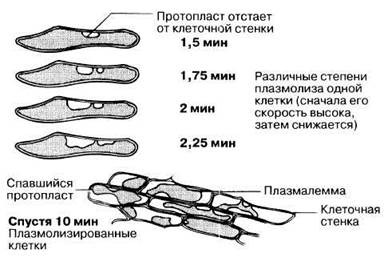
Fig. 13.3. Morphological changes in onion epidermal cells during plasmolysis. Epidermal strips were immersed in a 1 M sucrose solution for varying durations.
Last update: 06/08/2026
Editorial and Educational Adaptation: This material has been compiled based on the primary/original source text. The project team performed an editorial review, corrected technical inaccuracies, structured sections, and adapted the content for an educational format.
What was processed:
- elimination of formatting defects (OCR errors, structural breaks, corrupted characters);
- editorial organization of content;
- standardization of terminology in accordance with academic sources;
- verification of factual statements against the original source text.
All mentions of the author, publication year, and origin of the primary text have been preserved in accordance with the source.