BIOLOGY Volume 2 - A Guide to General Biology - 2004
18. THE ANIMAL SKELETOMUSCULAR SYSTEM
18.6. Locomotion in Vertebrates
18.6.5. Human Locomotion
Humans are characterized by bipedal locomotion, i.e., walking on two legs. Before examining this in more detail, it is helpful to review some of the concepts introduced in Section 18.4.1, specifically Muscle origin and insertion, flexors and extensors, and antagonistic Muscles.
Walking
A standing person maintains balance by supporting themselves on both legs. When taking a step forward with the right leg, the heel is first lifted off the ground As a result of the contraction of the gastrocnemius muscle; at the same time, the toes of the right FOOT push off the ground, generating forward propulsion. Next, the right leg is swung forward, bending slightly at the knee (Fig. 18.30). Body weight is transferred to the left leg, which remains on the ground and serves as body support. As the right leg straightens, the heel touches the ground first. Body weight is gradually shifted from the left foot to the right heel, and then to the big toe of the right foot. Typically, the big toe of one leg or the other presses against the ground at the end of each step, just as it pushes off at the beginning of the next. Now that the body weight has been transferred to the right leg, the left heel lifts, and the entire sequence of events repeats.
Class="center">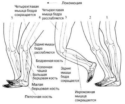
Fig. 18.30. Successive positions of the human right leg during a single step.
Antagonistic Muscles
The muscle mechanics involved in walking are quite complex, involving coordinated contractions and relaxations of many antagonistic muscles. The principles of their interaction can be illustrated by the flexion and extension of the leg at the knee joint. This joint is hinge-like, meaning movements occur in only one plane. The flexors in this case are the muscles at the back of the thigh. One of these is the biceps femoris, which simultaneously acts as an extensor at the hip joint, i.e., it participates in moving the thigh away from the torso. The extensors run along the front of the thigh. The main one is the quadriceps femoris. It straightens the leg at the knee joint and simultaneously flexes it at the hip, pulling the knee toward the torso. The origins and insertions of these muscles are attached to the skeletal structures as follows (Fig. 18.31).
1. Biceps femoris:
Origin → Pelvic Girdle and upper Femur
Insertion → Upper PARTS OF THE Tibia and Fibula
2. Quadriceps femoris:
Origin → Pelvic girdle and upper femur
Insertion → Patella
These muscles work antagonistically, meaning when one contracts, the other relaxes. As a result, the movements indicated by the arrows in Fig. 18.31 take place. They are triggered by motor neuron impulses reaching the muscle fibers and are coordinated by the Reflexes described below.

Fig. 18.31. Example of an antagonistic muscle pair involved in walking.
Inhibitory Reflexes
To move a limb back and forth, it requires at least a pair of antagonistic muscles: when one contracts, the other must relax. This is achieved by a simple inhibitory mechanism. Typically, when a postural muscle begins to stretch under an external load, the stretch receptors within it (a type of proprioceptor known as muscle spindles) respond to the elongation. They send nerve impulses to the Spinal Cord, and from there a signal travels via a motor neuron to trigger a counter-contraction that resists The change in body position. Imagine, for example, that a weight is placed on the palm of your outstretched arm. The arm begins to lower under the weight, and the biceps brachii stretches involuntarily. However, the contraction force reflexively increases, the increased load is balanced, and the arm remains in its previous position. This is an unconditioned reflex, but if necessary, it can be brought under conscious control. If you straighten your arm at the elbow joint, the extensor on the back of the arm contracts, and the biceps stretches. It is important to inhibit its normal reaction that would counteract your movement.
The sensory neuron running from the stretch receptor to the spinal cord is synaptically connected to interneurons in the Gray matter of the spinal cord (Fig. 18.32). Upon appropriate stimulation, they inhibit the excitation of motor Neurons innervating the antagonistic muscle, with the result that this muscle remains relaxed.

Fig. 18.32. Reciprocal inhibition. When muscle A (e.g., the biceps femoris) contracts, nerve impulses travel from it to the spinal cord, exciting an inhibitory interneuron. The contraction of muscle A requires the simultaneous relaxation and stretching of muscle B (e.g., the quadriceps femoris). This stimulates stretch receptors in the latter, which send impulses to the spinal cord. If the inhibitory interneuron did not exist, these impulses would excite the motor neuron driving the contraction of muscle B. When muscle B contracts, a similar system causes muscle A to relax.
A good example of this mechanism in action is walking. When the leg begins to bend at the knee as it leaves the ground, the extensors are still stretched, and stretch receptors reflexively inhibit their contraction. Then the leg straightens and touches the ground again. The flexor muscle no longer contracts, inhibition of the extensor muscle ceases, and stretching occurs, causing the extensor muscle to contract. When the leg is straight, the extensor muscle receptor receives no signal, and no stretch reflex is observed. The described cycle repeats with every new step.
18.6. Why do sprinters typically run on their toes?
Last update: 06/08/2026
Editorial and Educational Adaptation: This material has been compiled based on the primary/original source text. The project team performed an editorial review, corrected technical inaccuracies, structured sections, and adapted the content for an educational format.
What was processed:
- elimination of formatting defects (OCR errors, structural breaks, corrupted characters);
- editorial organization of content;
- standardization of terminology in accordance with academic sources;
- verification of factual statements against the original source text.
All mentions of the author, publication year, and origin of the primary text have been preserved in accordance with the source.