BIOCHEMISTRY - L. Stryer - 1984
VOLUME 3
Part IV INFORMATION
CHAPTER 25. RNA INFORMATION AND TRANSCRIPTION
25.15. Template DNA Contains Transcription Stop Signals
METABOLISM/31.html">Transcription termination is regulated just as finely as initiation. Template DNA contains stop signals that have been deciphered by comparing various base sequences in these regions. They all share a common feature: a GC-rich region followed by an AT-rich sequence just upstream of the termination site. A hallmark of termination sequences is the twofold Symmetry of this GC-rich region (Fig. 25.17). Consequently, the RNA transcript of this region is self-complementary, meaning it is capable of base-pairing to form a hairpin Structure (Fig. 25.18). In addition, the newly synthesized RNA chains end with several U residues, which are encoded by a run of A bases in the AT-rich region of the DNA template. One or more of these structural features cause RNA polymerase to stall and pause when it encounters such a signal. At some termination sites, newly formed RNA chains are released without the involvement of additional Proteins. At other sites, the participation of the p (rho) protein is required for chain termination.
Class="center">Fig. 25.17. DNA sequence corresponding to the 3' end of the E. coli trp mRNA. The base sequences shown in yellow exhibit twofold symmetry about the axis indicated in green.
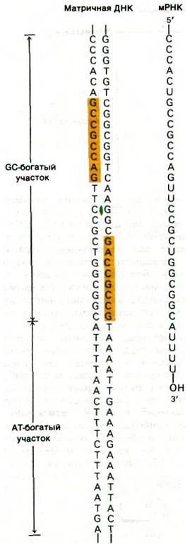
Fig. 25.18. Base sequence of the 3' end of mRNA transcribed from the E. coli Tryptophan Operon. A stable hairpin structure can potentially form

25.16. The p Protein Participates in Transcription Termination
The fact that transcription termination at certain sites involves the p protein is supported by the following evidence: RNA molecules synthesized in vitro in the presence of the p protein are shorter than those obtained in its absence. For example, RNA synthesized on fd phage DNA in the presence of the p protein has a sedimentation coefficient of 10S, whereas RNA synthesized in the absence of the p protein is 23S. Further insights into the action of the p protein were gained by adding this termination factor to the incubation mixture at various time intervals after the onset of RNA Synthesis. When the p protein was added a few seconds, 2 min, and 10 min after initiation, RNAs with sedimentation coefficients of 13S, 17S, and 23S, respectively, were obtained. This result indicates that the template contains at least three p-sensitive termination sites (yielding 10S, 13S, and 17S RNA) and one p-independent termination site (yielding 23S RNA). Therefore, specific termination can occur even in the absence of the p protein. However, the p protein reveals additional termination signals that RNA polymerase cannot recognize on its own.
Fig. 25.19. Effect of the p factor on the size of transcribed RNAs

How does the p protein cause termination of RNA synthesis upon reaching specific termination signals? One possibility is that this 200-kDa tetrameric protein binds to the RNA and moves toward the RNA polymerase molecule, halting it at the termination site. According to this model, the p protein displaces RNA polymerase from the 3' end of the RNA, resulting in the release of the RNA transcript. Interestingly, the p protein hydrolyzes ATP during transcription termination.
As might be expected, the termination of transcription of certain genes is regulated. As we will see further on, bacteriophage  synthesizes anti-terminator proteins that allow the transcription and expression of certain genes (Sec. 28.11). E. coli employs a regulatory system using specialized termination signals called attenuators to meet The Cell's nutritional demands (Sec. 28.9).
25.17. Many RNA Molecules Are Cleaved and Chemically Modified After Transcription
The formation of functionally active RNA molecules (Processing) continues after transcription is complete. In prokaryotes, transfer and ribosomal RNA molecules are generated by the Cleavage and chemical modification of specific newly synthesized RNA chains. For example, in E. coli, Three types of ribosomal RNA molecules and one Transfer RNA molecule are excised from a primary RNA transcript that also contains spacer regions (Fig. 25.20). Other transcripts contain several Different types of tRNA or multiple copies of the same tRNA. The Nucleases that cleave and shorten these rRNA and tRNA precursors act with high precision. For instance, Ribonuclease P generates the correct 5' ends of all tRNA molecules in the E. coli cell. Ribonuclease III excises the 5S, 16S, and 23S rRNA precursors from the primary transcript by cleaving specific bonds in double-helical hairpin regions. By contrast, prokaryotic mRNA molecules undergo virtually no modification. Furthermore, many of them are translated even before their transcription is finished. At the same time, certain viral mRNAs (e.g., T7 phage mRNA) are cleaved by ribonuclease III before Translation begins.
A second type of processing is The addition of NUCLEOTIDES to the ends of certain RNAs. For example, the CCA sequence is added to the 3' ends of tRNA molecules that lack this terminal sequence. In eukaryotes, a long poly(A) sequence is added to the 3' end of most mRNA molecules, and a methylated G nucleotide (the so-called "cap") is added to the 5' end (Sec. 29.22).
Fig. 25.20. Cleavage of this primary transcript yields 5S, 16S, and 23S rRNA molecules and one tRNA molecule. Spacer regions are highlighted in yellow

Modifications of bases and ribose residues represent a third type of processing. In eukaryotes, roughly one out of every hundred ribose residues in rRNA has its 2'-hydroxyl group enzymatically methylated by S-adenosylmethionine. In Bacteria, it is the bases rather than the ribose residues that are methylated. Particularly interesting are the unusual bases found in all tRNA molecules (Sec. 27.3). They are formed by the enzymatic modification of ordinary ribonucleotides contained within the tRNA precursor. For example, pseudouridylate and ribothymidylate are formed by the post-transcriptional modification of uridylate residues.


In eukaryotes, all transcripts undergo extensive processing. The cleavage and modification of rRNA and tRNA precursors closely resemble the corresponding processes in prokaryotes. A striking difference is that eukaryotic mRNA is produced by the cleavage of a large transcript followed by the joining (splicing) of the resulting fragments. We will examine this phenomenon in a subsequent chapter (Sec. 29.21). In eukaryotes, transcription and translation take place in separate cellular compartments. RNA Processing likely plays a key role in regulating The transport of mRNA, rRNA, and tRNA from The Nucleus to the Cytosol.
Last update: 06/08/2026
Editorial and Educational Adaptation: This material has been compiled based on the primary/original source text. The project team performed an editorial review, corrected technical inaccuracies, structured sections, and adapted the content for an educational format.
What was processed:
- elimination of formatting defects (OCR errors, structural breaks, corrupted characters);
- editorial organization of content;
- standardization of terminology in accordance with academic sources;
- verification of factual statements against the original source text.
All mentions of the author, publication year, and origin of the primary text have been preserved in accordance with the source.