Biochemistry - The Chemical Reactions of Living Cells Volume 3 - D. Metzler 1980
Light in Biology
Photosynthesis
Pigments and Their Environment
Chlorophylls (Fig. 13-19) are structurally similar to Hemes (Fig. 10-1), but ring IV, unlike the corresponding ring D in Porphyrins, is not fully dehydrogenated. In addition, the chlorin ring Structure in chlorophyll is modified by The formation of a fifth ring (V) containing a ketone group and a methyl ester. As can be seen, ring V is formed by the cross-linking of the propionic acid side chain of ring III and the carbon of the methene bridge. This ring system is known as a pheoporphyrin. The Nature of the peripheral substituents on the ring indicates a close relationship between chlorophylls and porphyrins. However, in most chlorophylls, one of the carboxyethyl groups is esterified with a long phytol chain. Chlorophyll a is the principal pigment in METABOLISM/14.html">Chloroplasts and the most important chromophore for Photosynthesis in green plants. Other forms of chlorophyll, alongside carotenoids and certain other pigments, function as accessory pigments and act as a sort of “light-harvesting” system. Their relative Abundance in the photosynthetic unit of spinach chloroplasts (Section 4) is given in Table 13-2.
Class="center">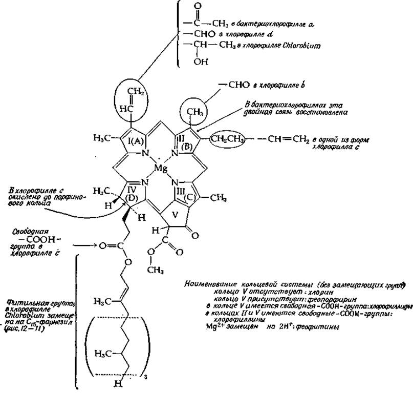
FIG. 13-19. Structural formulas of chlorophylls.
In 80% acetone, chlorophyll a exhibits an intense, narrow absorption band with Vmах = 663 nm (15,100 cm-1); in intact chloroplasts, this maximum is shifted toward the red region, with the bulk of the chlorophyll absorbing at 678 nm. Chlorophyll b is almost invariably present in green leaves (Fig. 13-19). Its absorption maximum in acetone is 635 nm (15,800 cm-1). Chlorophyll c is found in diatoms, brown Algae (Phaeophyta), and dinoflagellates (Figs. 1-7); it lacks the phytol group and is believed to be a mixture of two compounds. Chlorophyll d, which occurs alongside chlorophyll a in certain species of Rhodophyta (Chapter 1, Section D,3), has been only partially characterized [77].
Table 13-2 Approximate composition of an "averaged" photosynthetic unit of spinach chloroplastsa,b
|
Component |
Number of molecules |
|
Chlorophyll a |
160 |
|
Chlorophyll b |
70 |
|
Carotenoids |
48 |
|
Plastoquinone A |
16 |
|
Plastoquinone B |
8 |
|
Plastoquinone C |
4 |
|
a-Tocopherol |
10 |
|
a-Tocopherylquinone |
4 |
|
Vitamin K |
4 |
|
116 |
|
|
Sulfolipids |
48 |
|
Galactosylglycerides |
490 |
|
Iron |
12 atoms |
|
Ferredoxin |
5 |
|
Cytochrome b563 |
1 |
|
Cytochrome b559 |
|
|
Cytochrome f |
1 |
|
Copper |
6 atoms |
|
Plastocyanin |
1 |
|
Manganese |
2 atoms |
|
Protein |
928,000 daltons |
a From Gregory [77] [based on data by Lichtenthaler H. K., Park R. B., Nature (London), 198, 1070 (1963)] and White A., Handler P., Smith E. L., Principles of Biochemistry, 5th ed., p. 528, McGraw-Hill, New York, 1973.
b The number of molecules was calculated on the assumption that each unit contains two Mn2+ ions.
Photosynthetic Bacteria contain bacteriochlorophylls, in which ring II is reduced (Fig. 13-19). The absorption band of these compounds is shifted further into the red region relative to chlorophyll a, reaching approximately ~770 nm. The principal chlorophyll of green sulfur bacteria of the genus Chlorobium—chlorobium chlorophyll—features hydroxyethyl and farnesyl side chains. Pheophytins are derivatives of chlorophyll formed by the removal of Mg2+ upon Treatment with weak acid. Hydrolysis of the ester methyl group yields chlorophyllides, while the simultaneous removal of both the methyl and phytol groups yields chlorophyllins.
Because chlorophylls are readily and completely extracted by mild Solvents [81], one might assume they are simply dissolved in the lipid phase of the membranes. However, the absorption spectrum of chlorophyll in leaves exhibits bands shifted toward the red relative to those of chlorophyll a in acetone, with shifts reaching up to 900 cm-1. In most green plants, chlorophyll displays at least four main absorption bands with λmах = 662 nm (15,120 cm-1), 670 nm (14,940 cm-1), 677 nm (14,770 cm-1), and 683 nm (14,630 cm-1) [82]. Minor bands with vmax = 14,420 and 14,230 cm-1 are also occasionally observed (Fig. 13-20). This suggests that chlorophyll molecules reside in varying microenvironments within the membranes. As a result, the absorption spectrum broadens, facilitating more efficient light capture. It is believed that reaction centers also contain chlorophyll; in Photosystem I, it absorbs at ~700 nm (14,290 cm-1), whereas in Photosystem II, it absorbs at ~682 nm (14,660 cm-1).

FIG. 13-20. A. Absorption spectrum analysis of chlorophyll in a suspension of chloroplast fragments from Scenedesmus [82]. B. Schematic representation of the polypeptide backbone conformation and the arrangement of chlorophyll molecules in a subunit of the bacteriochlorophyll-protein complex [Fenna R. E., Matthews B. W., Nature (London), 258, 573–577 (1975)]. Circles indicate the presumed positions of a-carbon atoms. The course of the polypeptide chain remains undetermined in several regions (dashed lines). For clarity, magnesium atoms, chlorophyll ring substituents, and phytol chains (except the first one) are omitted. C. Proposed arrangement of a pair of chlorophyll a molecules in reaction centers [93a]. R = phytol; R' = a Water molecule hydrogen or an amino acid residue of the protein.
The bacteriochlorophyll found in Chromatium Cells also exhibits three absorption bands with λmах = 800, 850, and 890 nm. The latter band corresponds to the bacteriochlorophyll of the reaction center, which is the only form that fluoresces. A water-soluble bacteriochlorophyll-protein complex isolated from green photosynthetic bacteria of the genus Chlorobium has been successfully crystallized. X-ray crystallographic analysis of its three-dimensional structure [83] has demonstrated that each subunit (with a Molecular Weight of 50,000) of the trimeric molecule contains seven embedded bacteriochlorophyll molecules, as illustrated in Fig. 13-20,B. In green plants, chlorophyll may also exist in association with more hydrophobic Proteins. Two such complexes have been isolated [84], one of which is thought to belong to photosystem I and the other to photosystem II.
Irradiation of chloroplasts induces readily measurable chlorophyll a fluorescence, whereas chlorophyll b, other chlorophyll forms, carotenoids, and other pigments exhibit no fluorescence whatsoever. This indicates that all of these substances function as accessory pigments that efficiently transfer energy to the chlorophyll a located in the reaction center. As seen in Fig. 13-21, the absorption bands of accessory pigments generally lie in higher-energy regions than those of the reaction centers. Thus, the photosynthetic Organism absorbs light across a broad wavelength range, and all the captured energy is subsequently funneled into the reaction centers.

FIG. 13-21. Absorption spectra of chlorophylls and accessory pigments [Govindjee G. and R., Sci. Am., 231, 68–82 (Dec. 1974)].
Among the most important accessory pigments are the carotenes (Fig. 12-14), of which ß-carotene is the principal representative in most green plants. Green sulfur bacteria contain y-carotene; one end of this molecule remains uncyclized and resembles lycopene. Chloroplasts contain a variety of oxygenated carotenoids (xanthophylls). Among these, neoxanthin, violaxanthin [Eq. (12-30)], and lutein predominate in Higher Plants and green algae. Lutein resembles zeaxanthin, but the ring at one end of its chain undergoes isomerization via the migration of a double bond to the position shown below:

Euglena and related microorganisms contain high levels of antheraxanthin (Section D,7), whereas brown algae and diatoms are dominated by fucoxanthin and zeaxanthin (Fig. 12-14). Purple sulfur bacteria of the genus Rhodospirillum rubrum synthesize a distinct spirilloxanthin; both ends of this molecule feature the structure shown below:

Another, less widely distributed class of accessory pigments comprises open-chain tetrapyrroles [85]; due to their structural resemblance to Bile pigments (Fig. 14-14), they are frequently referred to as "plant bile pigments." Phycocyanins impart the characteristic color to blue-green algae. They form a group of Conjugated Proteins (biliproteins) containing phycocyanobilin as their covalently bound prosthetic group (Fig. 13-22). Similarly, red phycoerythrins from Rhodophyta contain bound phycoerythrobilin (Fig. 13-22). Although these pigments are sometimes described as linear tetrapyrroles, it is entirely possible that their molecules adopt a helical conformation in vivo (Fig. 13-22). Algal biliproteins (phycocyanins and phycoerythrins) apparently aggregate to form distinct granules located on the outer surface of photosynthetic membranes. In blue-green algae, these granules are known as phycobilisomes.
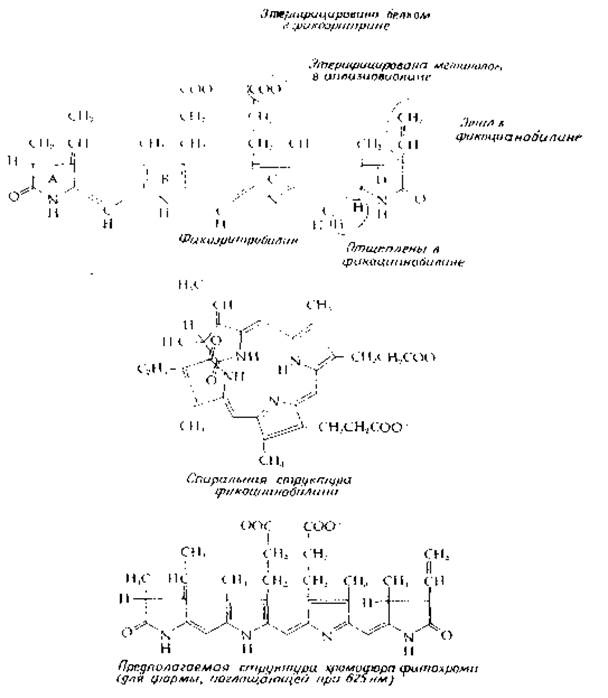
FIG. 13-22. Structures of open-chain tetrapyrroles found in plants (see also Fig. 14-14).
Last update: 06/08/2026
Editorial and Educational Adaptation: This material has been compiled based on the primary/original source text. The project team performed an editorial review, corrected technical inaccuracies, structured sections, and adapted the content for an educational format.
What was processed:
- elimination of formatting defects (OCR errors, structural breaks, corrupted characters);
- editorial organization of content;
- standardization of terminology in accordance with academic sources;
- verification of factual statements against the original source text.
All mentions of the author, publication year, and origin of the primary text have been preserved in accordance with the source.