Principles of Biochemistry Volume 1 - A. Lehninger 1985
Biomolecules
Vitamins and Trace Elements: Their Role in Enzyme Function
Vitamin A probably performs several functions
The Role of vitamin A as an essential dietary factor was first established in 1915 by Elmer McCollum, who later isolated this vitamin from fish Liver oil. Two natural forms of vitamin A are known: vitamin A1, or retinol, derived from marine fish liver, and vitamin A2, isolated from freshwater fish liver. Both of these Vitamins are 20-carbon alcohols composed of isoprene units. Vitamin A as such does not occur in plants, but many plants contain isoprenoid substances called carotenoids, which can be enzymatically converted into vitamin A in the bodies of most animals. Fig. 10-19 illustrates how vitamin A is formed from the Cleavage of ß-carotene, which gives carrots, sweet potatoes, and other vegetables their characteristic color.
In humans and experimental animals, Vitamin A deficiency leads to a wide range of symptoms that at first glance seem unrelated. These include dry Skin, xerophthalmia ("dry eyes"), dryness of mucous membranes, delayed GROWTH AND DEVELOPMENT, male sterility, and night blindness. The latter symptom is commonly used for the early Diagnosis of vitamin A deficiency (Sec. 26.16).
Intensive biochemical and biophysical research on vitamin A, initiated by George Wald at Harvard University, has provided comprehensive insight into its function in the visual process. Fig. 10-20 illustrates the cycle of chemical Changes in the visual pigment rhodopsin within retinal rod Cells. These cells detect low-intensity light signals but are insensitive to color. The active component in Vision is the oxidized form of retinol—retinal, or vitamin A aldehyde—bound to the protein opsin. The retinal-opsin complex, known as rhodopsin, is located in stacked intracellular membranes of the rods. Upon excitation of rhodopsin by visible light, retinal—in which the double bond at the 11-position is in the cis-configuration (with all other double bonds in the trans-configuration)—undergoes a series of highly complex and rapid intramolecular rearrangements, isomerizing to all-trans-retinal. These changes, which alter the geometric configuration of retinal (Fig. 10-20), are believed to induce a conformational change in the entire rhodopsin molecule. This conformational shift acts as a molecular trigger that generates an impulse in the Optic nerve endings, which is then transmitted to the Brain. Through subsequent "dark" enzymatic reactions, the light-induced all-trans-retinal is converted back into the original 11-cis-retinal.
Class="center">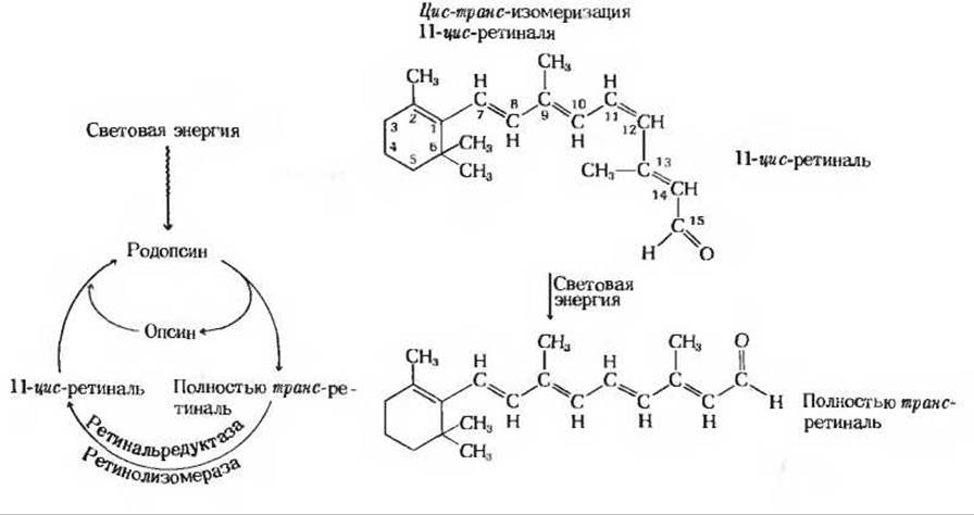
Fig. 10-20. The cyclic process of Synthesis and Breakdown of the visual pigment rhodopsin. Upon excitation of a rhodopsin molecule by visible light, its prosthetic group, 11-cis-retinal, absorbs light energy and undergoes a multistep isomerization into all-trans-retinal, a process that triggers a Nerve Impulse. Because The Structure of all-trans-retinal does not fit the conformation of the Active Site of the opsin protein, it dissociates from the protein. Through two successive enzymatic reactions, all-trans-retinal is converted back into the original 11-cis-retinal, which then rebinds to opsin to regenerate rhodopsin.
Retinal is also found in Bacteriorhodopsin, a light-sensitive pigment consisting of a retinal-protein complex present in The Cell membrane of halobacteria—"salt-loving" prokaryotes that derive most of their energy from light absorbed by this pigment (Ch. 17).
Last update: 06/08/2026
Editorial and Educational Adaptation: This material has been compiled based on the primary/original source text. The project team performed an editorial review, corrected technical inaccuracies, structured sections, and adapted the content for an educational format.
What was processed:
- elimination of formatting defects (OCR errors, structural breaks, corrupted characters);
- editorial organization of content;
- standardization of terminology in accordance with academic sources;
- verification of factual statements against the original source text.
All mentions of the author, publication year, and origin of the primary text have been preserved in accordance with the source.