Chemistry and Biology of Proteins - F. Haurowitz 1953
Proteins with Enzymatic Properties
Flavoproteins and Pyridine Nucleotides
The first yellow enzyme discovered in Yeast by Warburg and Christian [139] was isolated in crystalline form by Theorell [140]. This enzyme is extracted from yeast with Water and precipitated from the aqueous extract using acetone or methanol. Dialysis against dilute Hydrochloric acid or Treatment with high concentrations of methanol causes the yellow active group to dissociate from the enzyme. Hydrochloric acid denatures the protein component of the enzyme, as indicated by an increase in the number of free sulfhydryl groups; upon removal of the hydrochloric acid by dialysis, the protein (apoenzyme) renatures and regains its ability to bind with the yellow active group [140]. It was previously assumed that this group was riboflavin phosphate.
Class="center">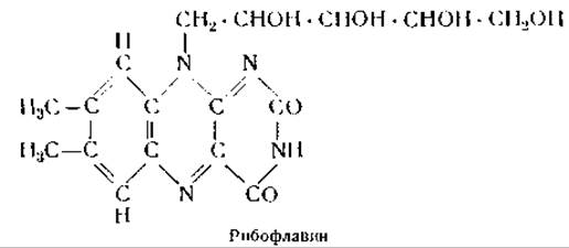
Later, however, it was found that the prosthetic group of Flavoproteins is flavin adenine dinucleotide, The Structure of which can be represented schematically by the formula Fl—F—P—R—Ad, where Fl is riboflavin, F is phosphoric acid, R is ribose, and Ad is adenine [139]. Despite the fact that flavoproteins are quite stable Enzymes, their colorless apoenzymes in the free state are extremely labile and become stable only upon binding with the prosthetic group [141]. The Molecular Weight of the yellow enzyme is approximately 80,000 [142], and each enzyme molecule contains 1 molecule of flavin adenine dinucleotide. It is highly probable that the dinucleotide is linked to the basic groups of the protein component via phosphoric acid.

The catalytic action of flavoproteins is based on the ability of their prosthetic groups to accept electrons and transfer them to other substances. The reduced form of the prosthetic group, designated as leucoflavin, is a colorless compound; however, upon giving up two electrons (i.e., becoming oxidized), it regains its yellow color.
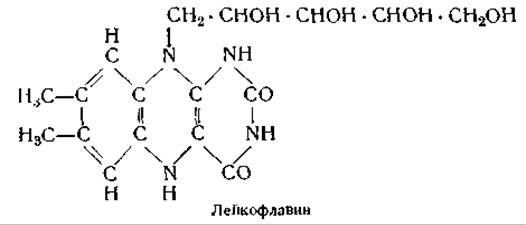
The profound Influence of the apoenzyme on the catalytic activity of the prosthetic group is confirmed by a significant shift in the oxidation-reduction potential of the flavin upon its combination with the protein; while the oxidation-reduction potential of a system consisting of equivalent concentrations of oxidized and reduced flavin is —0.060 V at pH 7, the oxidation-reduction potential of a system consisting of equivalent concentrations of oxidized and reduced flavoprotein is approximately —0.185 V [143].
The great importance of the protein component in flavoproteins is also manifested in the distinct Specificity of these enzymes. Currently, at least 10 different flavoproteins are known that contain either riboflavin phosphate or flavin adenine dinucleotide as a prosthetic group, yet differ from one another in their protein components. The most important representatives of flavoproteins are D-Amino Acid Oxidase [144], L-Amino Acid Oxidase, Glycine Oxidase [145], mold glucose oxidase [146], xanthine oxidase [147], fumaric acid Hydrogenase [148], cytochrome reductase [149], histaminase [150], and diaphorase [151]. The latter enzyme catalyzes the Oxidation of reduced pyridine NUCLEOTIDES (see below) in the presence of methylene blue. As already noted, various flavin enzymes differ from one another solely in the structure of their apoenzyme, although the exact nature of these differences has not yet been established. It also remains unknown how the apoenzyme affects the prosthetic group to bring about the specific catalysis of The oxidation of only certain types of compounds.
In this respect, flavoproteins are similar to enzymes that contain diphosphopyridine nucleotide (DPN) or triphosphopyridine nucleotide (TPN) as a prosthetic group [152]. There is, however, one essential difference between this group of enzymes and flavoproteins. Flavoproteins belong to the class of Conjugated Proteins whose non-protein components are bound more or less firmly to their specific proteins and are dissociated only under METABOLISM/18.html">The Influence of acid or methyl alcohol, whereas the apoenzymes of DPN and TPN are less tightly bound to their enzymes [153], such that even in neutral aqueous solutions, a large portion of the nucleotide remains in a free state. The Chemical Structure of DPN and TPN can be represented as follows: N—P—F—P—R—Ad and N—P—F—P—F—P—R—Ad, where N is nicotinamide, R is ribose, F is phosphoric acid, and Ad is adenine. Diphosphopyridine nucleotide, also called cozymase or cohydrase I, is extracted from yeast with hydrochloric acid [154]; after precipitation of the proteins with lead acetate and subsequent removal of lead from the filtrate, the nucleotide is adsorbed onto Norit and eluted with amyl alcohol and water [155]. Triphosphopyridine nucleotide was extracted from erythrocytes with acetone and precipitated from the acetone extract with mercuric acetate [156]. These two Coenzymes, by entering into temporary and loose combinations with specific proteins, form various enzymes [154].
The most important substrates for the action of enzymes containing DPN and TPN are lactic acid, triose phosphate, glucose, alcohol, glucose-6-phosphate, glycerophosphate, malic acid, isocitric acid, phosphogluconic acid, glutamic acid, and formic acid. As in the case of flavoproteins, the apoenzymes of various enzymes containing DPN and TPN are strictly specific; some apoenzymes have been obtained in crystalline form.
Alcohol dehydrogenase was obtained in crystalline form from yeast extract by precipitation with acetone or ethyl alcohol [157]. Lactic dehydrogenase was isolated from Heart Muscle, and its purification involved adsorption on tricalcium phosphate followed by elution at pH 7.2 [158]. A crystalline preparation of this enzyme was also obtained by precipitation with mercury salts followed by removal of the mercury through dialysis against a cyanide solution [159]. 1,3-Phosphoglyceraldehyde dehydrogenase, which catalyzes the oxidation of 1,3-phosphoglyceraldehyde to phosphoglyceric acid, was isolated in crystalline form from yeast juice by precipitation with nucleic acid; the latter was removed by precipitation with the protamine sturin, and subsequently, by carefully adding ammonium sulfate to the protein solution, enzyme crystals were obtained [160]. Crystalline 3-phosphoglyceraldehyde dehydrogenase was obtained from muscle by extraction with 0.03 N potassium hydroxide solution followed by precipitation with ammonium sulfate [161]. This enzyme was found in the myogen fraction of muscle (see Chapter VIII, p. 186), accounting for approximately 10% of the soluble Muscle Proteins [162]. The apoenzymes of phosphoglyceraldehyde dehydrogenase isolated from yeast and from muscle proved to be different [163]. The protein components of this entire group of enzymes likely possess not only substrate specificity but also species specificity. Pyridine enzymes also include cytochrome reductases, which reduce the ferric iron in cytochrome c to the ferrous state. It was found that cytochrome reductase from yeast has diphosphopyridine nucleotide as its prosthetic group, whereas the prosthetic group of Liver cytochrome reductase is triphosphopyridine nucleotide [164].
Last update: 06/08/2026
Editorial and Educational Adaptation: This material has been compiled based on the primary/original source text. The project team performed an editorial review, corrected technical inaccuracies, structured sections, and adapted the content for an educational format.
What was processed:
- elimination of formatting defects (OCR errors, structural breaks, corrupted characters);
- editorial organization of content;
- standardization of terminology in accordance with academic sources;
- verification of factual statements against the original source text.
All mentions of the author, publication year, and origin of the primary text have been preserved in accordance with the source.