Practical Protein Chemistry - A. Darbre 1989
Application of electron impact mass spectrometry for determining the amino acid sequence of peptides and proteins
General principles of mass spectrometric determination of the amino acid sequence of peptides and proteins
The main advantage of mass spectrometric amino acid sequencing lies in the possibility of analyzing peptide mixtures [24, 25]; otherwise, mass spectrometry could not compete with classical Methods for establishing the Introduction/19.html">Primary Structure of Peptides and Proteins, as its use is far more expensive than the manual dansyl-Edman Degradation method, which sometimes offers higher sensitivity. Protein chemists are well aware that the isolation of individual peptides from the mixture generated by enzymatic Protein Cleavage is precisely the rate-limiting step in determining peptide Amino acid sequences by classical techniques. Although the primary structure of large peptides is now routinely determined automatically using sequencers, locating these fragments within the protein chain always requires deeper, non-Specific Cleavage of the protein molecule to yield numerous small peptides. To determine the Amino Acid Sequence of these short peptides manually, the mixture must first be separated into individual components. When mass spectrometry is employed for structure determination, the isolation of individual components is completely unnecessary, and in the case of complex mixtures, only a crude fractionation of the mixture is required.
After modification via the method described above, the fraction under investigation is placed into the vaporizer and introduced into the ion source using a insertion rod. Due to the differing volatility of the modified peptides, individual components are fractionally sublimed from the mixture upon gradual heating; consequently, mass spectra recorded at specific time intervals typically characterize those peptides whose concentration in the gas phase is predominant. By comparing sample mass spectra obtained at various temperatures, one can unambiguously determine The amino acid sequence of individual peptides within the mixture. This approach yields excellent results when analyzing mixtures containing up to five different proteins. General strategies for establishing the Primary Structure of Proteins will be discussed below, enabling the most effective use of mass spectrometry advantages in sequencing short peptides. It should be borne in mind, however, that to achieve maximum efficiency when determining the primary structure of an unknown protein, classical and mass spectrometric methods should always be combined. Such an approach may proceed as follows:
1) the N-terminal amino acid sequence of the protein or large peptides is determined using automated sequencers;
2) peptides resulting from specific Enzymatic cleavage of the protein with Trypsin or Chymotrypsin are structurally analyzed using the manual dansyl-Edman method;
3) the amino acid sequence of short peptides—produced by non-specific Cleavage of the protein molecule with Elastase or subtilisin to determine the insertion sites of large peptides into the protein chain—is determined mass spectrometrically by analyzing their mixtures [2, 4, 25].
In A number of cases, mass spectrometry is the method of choice for solving structural problems. For instance, determining The nucleotide sequence of a DNA region corresponding to a specific Gene requires incomparably less effort if the partial amino acid sequence of the protein encoded by that gene has been determined by some method. Such information can be obtained within a few weeks using mass spectrometry. Mass spectrometry is an ideal method for rapidly assessing the degree of Homology among related proteins [4]. Furthermore, mass spectrometry is an unsurpassed technique for the structural analysis of amides and for determining THE POSITION OF Tryptophan residues within a peptide chain. The experimental protocol for amino acid sequencing using mass spectrometry can be outlined as follows:
1) the protein is cleaved non-specifically with elastase or subtilisin. The extent of Hydrolysis is verified by analytical Electrophoresis at pH 6.5;
2) the bulk of short peptides is separated from non-hydrolyzed protein and large peptides by Gel filtration;
3) the mixture of short peptides is separated by cation-exchange Column Chromatography (e.g., on Dowex [4, 25]) or High-Performance Liquid Chromatography. Optimal Separation on cation exchangers is achieved by elution with a pyridine-acetic acid mixture [4, 25], whereas in high-performance liquid chromatography, an acetic acid-propanol mixture is used [26]. The presence of peptides in the eluate is monitored by analytical electrophoresis (~ 1/100 of each fraction is used for analysis);
4) by selecting specific fractions and drying them, samples containing mixtures of up to five peptides are prepared;
5) the samples are chemically modified and analyzed by mass spectrometry.
Applying this approach allows an experienced researcher to obtain 70% of the amino acid sequence information for a protein with an M≤20,000 in approximately two months.
Class="center">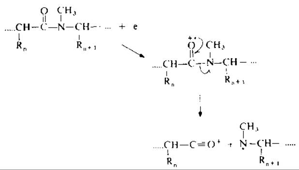
FIG. 19.3. C—N cleavage of peptide bonds in permethylated peptides.
Last update: 06/08/2026
Editorial and Educational Adaptation: This material has been compiled based on the primary/original source text. The project team performed an editorial review, corrected technical inaccuracies, structured sections, and adapted the content for an educational format.
What was processed:
- elimination of formatting defects (OCR errors, structural breaks, corrupted characters);
- editorial organization of content;
- standardization of terminology in accordance with academic sources;
- verification of factual statements against the original source text.
All mentions of the author, publication year, and origin of the primary text have been preserved in accordance with the source.