Protein Chemistry - Part 2 - Selected Topics in Special Protein Chemistry - Ashmarin, I. P. 1968
Histones
Isolation and Fractionation of Histones
Depending on the nuclear material content, Histones can be isolated either from pre-isolated nuclei or from whole Tissues. From the Thymus, where the nuclear content is high (up to 10% of dry weight), histones can be obtained in sufficiently large quantities if soluble cytoplasmic components are first removed by repeated washing—for example, with a 0.14 M sodium chloride solution acidified to pH 4. This approach makes it possible to eliminate the majority of non-histone Proteins and prevent histone aggregation, which reaches its maximum at pH above 4.5. For all other tissue groups, obtaining clean nuclei is essential. The most convenient Methods for their isolation are sucrose gradient centrifugation followed by washing and final purification According to the Chauveau and Moulé method. Washing the nuclei with citrate and acetate should be avoided, as this leads to the loss of a portion of Lysine-rich histones. Although isolating histones from nuclei yields purer preparations, it involves a significant loss of material and takes more time.
In addition to nuclei, nuclear deoxyribonucleoproteins (DNP) can be used to isolate histones. The latter can be obtained relatively easily by repeatedly washing the suspended tissue with a 0.14 M sodium chloride solution or a 0.075 M sodium chloride solution in 0.024 M EDTA, extracting DNP from the precipitate with a 1 M sodium chloride solution, and reprecipitating it in a 6-fold volume of distilled Water. More complex Procedures for obtaining DNP also exist, which include Chromatin isolation, water extraction of Nucleoproteins, and their purification from insoluble complexes by centrifugation.
To obtain total histone preparations, nuclei or DNP are extracted with 0.2–0.3 N hydrochloric or sulfuric acid, or with a mixture of 80% ethanol and 0.25 N Hydrochloric acid. Histones can be precipitated from acid extracts by adding ammonia up to pH 11, as well as by adding 5–10 volumes of ethyl alcohol or acetone. Most of these methods do not prevent histone loss and preparation contamination by acidic proteins and tissue proteases that are very similar in properties to Chymotrypsin. During isolation, the latter cleave histones, which is revealed by distorted electrophoretic Separation patterns in starch or polyacrylamide gels and the appearance of additional N-terminal amino groups. Histone degradation increases if they are extracted from pre-frozen tissues, because freezing and subsequent thawing cause proteases to penetrate various PARTS OF THE Cell and subsequently co-extract with histones. Tissue proteases are absent in nuclei, are inactive in acidic environments, and are easily inactivated by diisopropyl fluorophosphate. Therefore, the main conditions for successful histone isolation are treating the tissue at 0–2°C immediately after the animal's death, obtaining nuclei or DNP, using mildly acidic solutions to remove ballast proteins, and ensuring rapid extraction. The addition of diisopropyl fluorophosphate can be very useful. The histone yield varies considerably across different tissues, ranging from 0.12 to 2.4% of the wet tissue weight (see Table 7).
In the earliest works on histone fractionation, these proteins were successfully divided into two major groups: lysine-rich histones and Arginine-rich histones. They differed significantly in their Amino Acid Composition and physicochemical properties. For instance, the sedimentation constants for lysine-rich histones were 0.8–1.8S, and their molecular weights ranged from 8,000 to 20,000. For arginine-rich histones, these values were 1.5–2.6S and 37,000–51,000, respectively. Lysine-rich histones did not aggregate even at their isoelectric point, whereas arginine-rich histones readily formed aggregates upon an increase in pH, Ionic strength, buffer anion valence, and protein concentration.
The Development of chemical and physicochemical Fractionation Methods demonstrated that histones are complex, multicomponent compounds that can be subdivided into at least three main groups: 1) lysine-rich histones (fraction f1), 2) moderately lysine-rich histones (fraction f2), and 3) arginine-rich histones (fraction f3). These groups differ from one another in chemical and physicochemical parameters (amino acid composition, N-terminal groups, electrophoretic mobility, sedimentation constants), and above all in their lysine/arginine ratio, given that the sum of these Two Amino Acids is approximately the same across all fractions. For f1, this ratio is 10–13; for f2, it ranges from 1 to 2; and for f3, it is less than 1. Each of the listed groups is highly heterogeneous and can be further separated into several subfractions.
The most common techniques for histone separation include differential extraction, fractional precipitation, Column Chromatography, and starch or Polyacrylamide gel Electrophoresis. For example, differential extraction with a mixture of 1.25 N hydrochloric acid and 80% ethanol (4:1) can be used to isolate the arginine-rich fraction and a portion of the moderately lysine-rich histones. Lysine-rich histones and the remainder of the moderately lysine-rich histones are extracted from the precipitate using 0.25 N hydrochloric acid. A convenient method for extracting the f1 fraction is its direct extraction from the thymus or nuclei (DNP) of other tissues using 5% perchloric acid, followed by precipitation with trichloroacetic acid at a final concentration of 18%. An example of fractional precipitation is the isolation of arginine-rich and moderately lysine-rich histones from an acidified alcohol extract using 5 volumes of acetone or by adding ammonia to pH 11. Lysine-rich histones can be precipitated from acidic
solutions by adding 2–3 volumes of ethyl alcohol while alkalinizing the solution to pH 6.0. To precipitate arginine-rich histones from an acidic alcohol extract, dialysis against absolute alcohol can also be used, leaving moderately lysine-rich histones in solution.
Differential extraction and fractional precipitation techniques are usually combined into a unified chemical fractionation scheme. An example of such a scheme is the Johns method, which enabled the successful separation of calf thymus histones into four fractions: lysine-rich histones f1, moderately lysine-rich histones f2a and f2b, and arginine-rich histones f3.
The lysine/arginine ratios for fractions f2a and f2b were 1 and 2, respectively. In calf thymus histones, fraction f1 accounted for about 20% of the protein, fractions f2a and f2b accounted for 35% and 25%, respectively, and the arginine-rich histones f3 accounted for about 20%. Subsequently, the moderately lysine-rich histones f2a were separated into two subfractions (f2a1 and f2a2), bringing the total number of major histone fractions to five.
According to A number of data, acid extraction and fractionation of histones yield better results than alcohol methods. This is explained by the fact that acids themselves do not denature histones and protect them from Cleavage by tissue proteases. Alcohols, on the other hand, lead to partial Denaturation of histones, as evidenced by the transition of a portion of the protein from the native a-form to the ß-form. At the same time, alcohol fractionation makes it possible to obtain purer histone preparations because the solubility of non-histone proteins in an acidified alcohol solution is low. These drawbacks inherent to both chemical fractionation approaches can be circumvented to a certain extent by using Ion-exchange chromatography on Amberlite IRC-50 and carboxymethylcellulose (CMC) for histone separation.
The cation exchanger IRC-50 was first used for histone separation by Crampton et al. back in 1955. True, the number of isolated fractions did not exceed two, and protein sorption on the column reached 60%. A finer separation of total histones and almost complete recovery of material from the column were achieved when the sodium form of the resin and gradient elution with guanidine hydrochloride were introduced (Fig. 14); elution began at pH 6.8 and a guanidine hydrochloride concentration of 7–8%, which increased linearly to 40%. Under these conditions, total histones from the thymus and other tissues were separated into six main components. Lysine-rich histones were eluted from the column first as two partially overlapping peaks, Ia and Ib. Next, moderately lysine-rich histones were eluted as overlapping peaks IIa and IIb. Based on its amino acid composition, peak IIb corresponded to fraction f2b, and peak IIa to fraction f2a2. The remaining peaks III and IV on the column were eluted by rapidly increasing the guanidine hydrochloride concentration to 40%. In terms of their amino acid composition, these histones approached fractions f3 and f2a, respectively. In several studies, researchers managed to further divide peak II into four components (IIaa, IIa, IIb, and IIc), thereby increasing the total number of histone fractions to eight.
Class="center">Scheme of chemical fractionation of calf thymus histones (Johns, 1964)


Fig. 14. Chromatographic separation of calf thymus histones on an Amberlite IRC-50 column in a guanidine hydrochloride gradient (Bonner et al., 1966). Column dimensions 0.6x55 cm, fraction volume — 2.5 ml
In early experiments on histone fractionation on Amberlite IRC-50, too large a proportion of the material remained tightly bound to the column matrix. Therefore, in 1956, Davison and Shooter attempted to separate calf thymus histones on CM-Cellulose. Using Buffer solutions with various pH values (from 3 to 6), they isolated a peak of lysine-rich histones and a peak consisting of a mixture of moderately lysine-rich and arginine-rich histones. In addition, minor peaks of acidic proteins and relatively lysine-rich histone aggregates were detected. To reduce aggregation effects, Phillips and Jones used sodium acetate buffer (pH 4.2) and dilute hydrochloric acid solutions (0.01–0.02 N) for histone elution, achieving the separation of thymus histones into 4 distinct fractions. The buffer at pH 4.2 eluted two peaks of lysine-rich histones IIa and IIb (with lysine/arginine ratios of 9 and 6, respectively), 0.01 N hydrochloric acid eluted moderately lysine-rich histones f2 (lysine/arginine = 1.3), and 0.02 N hydrochloric acid eluted arginine-rich histones f3 (lysine/arginine = 0.8).
In addition, a peak of non-histone proteins fx, presumably of an acidic nature, was detected (Fig. 15).
Ion-exchange chromatography methods are frequently combined with fractional extraction and precipitation techniques, making it possible to obtain 10 or more histone fractions (Table 8). For instance, lysine-rich histones f1 isolated from the thymus by the Johns method can be further resolved into three fractions by chromatography on CM-cellulose (eluted with borate buffer, pH 9, with stepwise increasing sodium chloride concentrations) and into four fractions by chromatography on IRC-50 (eluted with guanidine phosphate).
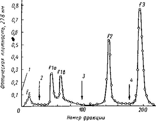
Fig. 15. Chromatographic separation of calf thymus histones on a carboxymethylcellulose column (Johns et al., 1960).
Column dimensions 14 x 2 cm, fraction volume — 3 ml. Stepwise elution with solutions: 1 — 0.1 M CH3COOH and 0.03 M NaOH; 2 — 0.17 M CH3COOH, 0.05 M NaOH, and 0.42 M NaCl; 3 — 0.01 N HCl; 4 — 0.02 N HCl
For finer resolution of "chemical" histone fractions, the countercurrent distribution method is also frequently employed; this technique has made it possible to identify seven subfractions within the lysine-rich histones f1 and four within the moderately lysine-rich histones f2b.
Last update: 06/08/2026
Editorial and Educational Adaptation: This material has been compiled based on the primary/original source text. The project team performed an editorial review, corrected technical inaccuracies, structured sections, and adapted the content for an educational format.
What was processed:
- elimination of formatting defects (OCR errors, structural breaks, corrupted characters);
- editorial organization of content;
- standardization of terminology in accordance with academic sources;
- verification of factual statements against the original source text.
All mentions of the author, publication year, and origin of the primary text have been preserved in accordance with the source.