Amino Acids, Peptides and Proteins - Dévényi T., Gergely J. 1976
Protein Analysis by Low-Voltage Electrophoresis
Low-Voltage Paper Electrophoresis
Low-Voltage Paper Electrophoresis
Principle of the method. Components of a protein mixture applied to a special filter paper moistened with a buffer solution migrate in an electric field in a direction and at a rate depending on their molecular charge. As a result, the mixture is separated into fractions that can be detected using specific staining Methods and quantified.
Applications: Qualitative and quantitative Analysis of Protein mixtures; qualitative and quantitative Analysis of Proteins in serum, urine, cerebrospinal, and tissue fluids.
Electrophoresis apparatus. A schematic diagram of the electrophoresis apparatus is shown in Fig. 1. It consists of a plastic chamber with a tight-fitting Glass lid 1. Both electrode chambers are divided into two compartments by a plastic partition 2. Platinum electrodes with a diameter of 0.5–0.8 mm are located in the outer compartments 3. The ends of the filter paper 4 are immersed into each of the two inner compartments. A connecting aperture is located at a height of 40 mm in the middle of the partition separating the outer and inner compartments of the electrode chamber. This aperture (0.5 cm in diameter) is loosely plugged with glass wool.
The electrophoresis chamber contains a removable plastic frame 5, on which filter paper strips are placed.
Power source. A regulated power supply provides a direct current of 100 mA and a voltage of up to 300 V.
Staining vessel. Filter paper strips are stained in a glass vessel with a lid measuring 7 x 38 cm.
Class="center">
Fig. 1. Schematic of the Grassmann-Hannig apparatus for paper electrophoresis [11]. See text for description.
PERFORMING ELECTROPHORESIS
1. Preparation of the buffer solution. Dissolve 29.43 g of veronal and 19.43 g of CH3COONa ∙ 3Н2О in approximately 1 L of distilled Water, then add 180 mL of 0.1 N НСl and bring the volume up to 3 L with distilled water. The buffer solution should have a pH of 8.6.
2. Filling the electrophoresis chamber with buffer solution. Pour approximately 500 ml of buffer solution into each compartment of the electrophoresis chamber. The buffer solution must reach the same level in all compartments of the apparatus.
3. Wetting the filter paper strips with the buffer solution. Before wetting, use a pencil to mark the position corresponding to the end of the frame and the Sample application site on the filter paper strip. The paper strips are then immersed and drawn through the buffer solution in the glass vessel with a smooth, steady motion. To submerge the strip, hold its ends with forceps, dip the right end into the buffer solution, and then slowly pull it upward while gradually submerging the left end. The strip should be moved at a speed that matches The rate of its wetting by the buffer. After wetting, remove the strip vertically from the buffer solution and allow the excess liquid to drain. Then, gently blot both sides of the strip with several layers of filter paper, applying only light contact to avoid squeezing out the moisture. Place the wet strips onto the frame According to the previously made marks.
4. Application of the test sample (serum). Pipette 0.01 ml of normal serum containing 5.6–8% protein onto the marked starting line of the pre-wetted strip.
Serum application method. The sample is applied with a pipette across the strip, leaving a 3 mm margin from each edge. To do this, the pipette is moved slowly and evenly along the starting line, its tip lightly touching the paper. The pipette tip must be smooth to avoid scratching the paper; a gentle Touch is sufficient for the sample to be absorbed.
5. Electrophoresis. The filter paper strip is placed on a frame inside the electrophoresis chamber so that both of its protruding ends are immersed in the buffer solution (Fig. 1). Once the strips are in place, the chamber is covered with a glass lid, and the apparatus is connected to the power supply. Electrophoresis is run for 12 to 16 h at 110 V and a current of approximately 1 mA per strip.
6. Removal and drying of paper electrophoregrams. Once the Separation of fractions is complete, the electric current is switched off and the glass lid of the apparatus is carefully lifted. Care must be taken to prevent Condensation accumulated on the inner surface of the lid from dripping onto the paper strips. The frame holding the strips is removed from the electrophoresis chamber and placed in a wire basket so that the ends of the strips, which previously lay near the edge of the frame, are now positioned on the edges of the basket. The basket with the strips is then placed in a drying oven at 110° C until the paper is dry. At this Temperature, the proteins denature and become fixed to the paper.
7. Staining of paper electrophoregrams. Protein fractions separated by electrophoresis are visualized on the paper strips by staining with Amido Black.
Staining solution. A cold-saturated solution of Amido Black is prepared in a solvent containing a mixture of glacial acetic acid and methanol (1:9). For this purpose, approximately 13,0 g of Amido Black is dissolved in 100 ml of acetic acid, and 900 ml of methanol is added. The mixture is thoroughly mixed by shaking and allowed to stand overnight; it is filtered the next day. The same portion of the stain can be reused several times.
Washing solution: acetic acid — methanol (1:9).
Staining procedure. The staining vessel is filled with the dye solution, and the heat-fixed paper electrophoregrams are immersed in it. Staining is performed for 10 min with continuous stirring, then the dye solution is replaced with a washing solution. The solution is changed several times over approximately 4 h until a fresh portion remains colorless.
NOTES: ELECTROPHORESIS CHAMBER
In addition to the Grassmann–Hannig apparatus, Other types of electrophoresis chambers are also used. Diagrams of two of these are shown in Figs. 2 and 3.
A. Durham chamber [6]. This apparatus is convenient for routine analyses (Fig. 2).
B. The Kunkel-Tiselius apparatus [18]. In this device, paper electrophoresis strips are placed between two siliconized plastic plates to eliminate unwanted evaporation of the buffer solution, thereby preventing changes in its concentration. Furthermore, this apparatus is also suitable for two-dimensional electrophoresis (Fig. 3).
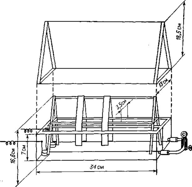
Fig. 2. Durham apparatus for paper electrophoresis [6].
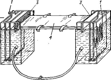
Fig. 3. Kinkel–Tiselius apparatus for paper electrophoresis [18].
1 — electrodes; 2 — glass wool layer in the slit between compartments; 3 — filter paper wick; 4 — plexiglass plate.
C. The "Elfor" chamber (Bender-Hobein, FRG). The plastic "Elfor" chamber is widely used for routine work in clinical laboratories. The apparatus ensures a constant pH value during electrophoresis. This is facilitated by a specially selected volume ratio of the electrode and analytical chambers, a specific method of laying paper strips, and an original labyrinth design around the electrodes. Laying the paper strips is very simple: they adhere on their own to the entire surface of the supporting removable bridge.
A. Buffer solutions used for serum Protein Electrophoresis must have a pH between 8.0 and 9.0. At these pH values, proteins migrate toward the anode (the isoelectric points of various Serum proteins range from pH 4.7 to 7.0).
B. The Ionic strength of the buffer solutions should be within 0.06–0.1 μ. Dilute solutions with an ionic strength below 0.06 μ lack sufficient buffering capacity and do not provide sharp separation of protein fractions.
C. From a practical standpoint, It is important to note that by increasing the voltage during electrophoresis, one can work with low ionic strength solutions. This will reduce heat generation under the Influence of the electric current, but the protein separation will not be very sharp. Increasing the ionic strength yields much better separation.
D. In addition to the buffer solutions mentioned above, the following can be used for protein and peptide electrophoresis:
1) Sodium barbiturate – barbituric acid buffer solution: 10.3 g of sodium barbiturate is dissolved in 1000 ml of distilled water, and 1.84 g of barbituric acid, also dissolved in 1000 ml of distilled water, is added; this yields a buffer solution with pH 8.6, μ = 0.06.
2) Sodium barbiturate – sodium acetate buffer solution: 7.36 g of Na barbiturate and 3.86 g of CH3COONa∙3H2О are dissolved in 1100 ml of distilled water; pH 9.0, μ = 0.06.
3) Borate buffer solution: 8.8 g of sodium borate and 4.05 g of boric acid are dissolved in 1000 ml of distilled water; pH 8.6.
4) Tris-EDTA buffer solution [1]. This solution is better suited than others for separating serum proteins. It allows the resolution of 9 fractions: prealbumin, albumin, three α-globulin fractions, three β-globulin fractions, and γ-globulin.
Preparation of the buffer solution. 60.5 g of tris(hydroxymethyl)aminomethane (Tris), 6.0 g of EDTA, and 4.6 g of boric acid are dissolved in distilled water and made up to a final volume of 1000 ml. The molarities of the solutes are 0.5, 0.021, and 0.075 M, respectively, and the pH is 8.9.
E. Buffer solutions are highly susceptible to mold or bacterial growth. Bacterial contamination should be carefully avoided, and contaminated solutions must not be used.
F. Buffer solutions should be stored in a refrigerator. It is impractical to prepare stock solutions in volumes exceeding weekly requirements, as their shelf life is no longer than 8–10 days. After each electrophoresis run, the polarity must be reversed, or the buffer solutions from the anode and cathode compartments must be thoroughly mixed in a bottle and poured back into the apparatus. Even when this requirement is met, the same portion of buffer solution should not be used more than 3–4 times.
SAMPLE APPLICATION
Two highly convenient methods for applying the sample to a strip of filter paper are described below.
A. Using a capillary pipette, the protein solution to be analyzed is transferred to the ground edge of a Microscope slide, then this edge of the slide is quickly applied to the paper strip and held until the entire solution is absorbed into the paper. Using a 2.5 cm wide slide, 0.05 ml of the test solution can be applied.
B. A U-shaped fork is made of plexiglass or another suitable plastic, with its prongs spaced 6–8 cm apart. Two platinum wires 0.1 mm thick are stretched between the ends of the prongs so that a gap of 0.5–1.0 mm remains between them. The space between the wires is filled with the test solution using a capillary pipette, and, after inverting the fork, the wires are quickly touched to The surface of the moistened paper strip.
VOLTAGE IN ELECTROPHORESIS
A. Under standard experimental conditions (the same paper, buffer solution, and apparatus), protein migration in an electric field depends on the voltage.
B. Typically, the recommended voltage of 110 V is used for serum protein fractionation, where separation takes 14 to 16 hours and no cooling is required. For special purposes, the voltage can be increased, but according to Joule's law, this will correspondingly increase heat generation. Heat generation can be reduced by lowering the ionic strength of the solution. If the filter paper used for electrophoresis is sufficiently moist and the ambient air temperature is not too high, the voltage across the strip can be increased to 250–300 V without taking special cooling measures. However, with any further increase in voltage, special cooling devices are required.
SOME FACTORS INFLUENCING ELECTROPHORESIS
A. Electroosmosis. When an electric current passes through a capillary system (filter paper) filled with a buffer solution, electroosmosis occurs, resulting in the movement of the buffer solution toward the cathode, counter to protein migration.
B. Factors affecting buffer solution flow:
1) After immersing the ends of the wet filter paper strip into the buffer solution filling the compartments of the electrophoresis apparatus, the solution begins to flow from one compartment to another along the wetted strips. This flow continues for 30 to 60 min until equilibrium is reached. If the paper strips were thoroughly pre-wetted, the equilibration period may be shorter.
To completely eliminate The Effect of this process on protein separation, the buffer-wetted paper strips should be placed on a frame, positioned inside the electrophoresis chamber, and the lid closed. Only after equilibrium is established, i.e., after approximately 30 min, can the sample be applied.
2) Joule heating increases the evaporation of the buffer solution from the paper surface. Evaporation leads to concentration of the buffer solution; the resulting increase in ionic strength raises its electrical conductivity, which in turn leads to further heating and evaporation of the solution. The evaporated buffer solution is replenished by the solution from the apparatus compartments via capillary action, initiating a flow from the ends of the strip toward its center. At one end of the strip, this flow aligns with protein migration, while at the other end, it drags them in the opposite direction.
In principle, the adverse effects and intensity of these counter-flows are minimal because a humid chamber environment is rapidly established within a sealed electrophoresis apparatus. Furthermore, some electrophoresis chambers are designed with an inlet port for introducing steam, allowing electrophoresis to begin in a vapor-saturated atmosphere.
As a result of buffer solution evaporation, water condenses and forms droplets on the inner surface of the glass lid covering the electrophoresis chamber. The risk of water droplets falling onto the electrophoretogram during the run or when removing the lid can be easily avoided by lining the inner surface of the lid with a sheet of filter paper.
3) If the buffer solution filling the compartments of the electrophoresis chamber is at different levels, it will flow along the paper strips from the higher-level compartment to the lower-level one. Depending on its direction, this buffer flow can either retard or accelerate protein migration.
To prevent siphoning, the buffer solution levels in all compartments of the electrophoresis chamber should be carefully leveled before inserting the paper strips. Electrophoresis apparatus compartments are typically equipped with drain Valves connected by a rubber hose. After filling the apparatus with buffer solution, the drain valves are opened (or, if there are no valves, the clamp is removed from the connecting rubber hose) to level the liquid.
An even simpler way to equalize the levels is by using a T- or Y-shaped tube filled with buffer solution, with rubber hoses attached to its ends. Two of its ends are immersed in the buffer compartments of the electrophoresis chamber, and the buffer solution is drawn in through the third outlet. Once the tube is filled with the solution, this outlet is sealed with a clamp.
4) Before working with a given electrophoresis apparatus, the optimal application point for the sample on the paper strip should be determined empirically to achieve the best separation.
SELECTING FILTER PAPER
The following commercial brands of electrophoresis filter paper have proven effective in practice: Schleicher & Schuell 2043a, Schleicher & Schuell 2043b, Whatman 1, Whatman 4, Munktell 20, Macherey-Nagel 214.
PROTEIN ADSORPTION ON FILTER PAPER
A. During electrophoresis, some proteins are adsorbed onto the filter paper. Protein adsorption is significant at acidic pH and low ionic strength, but it is also observed at pH 8.6. Adsorption occurs due to the interaction between positively charged protein molecules and negatively charged filter paper fibers. The degree of adsorption varies widely among different proteins; for example, serum Lipoproteins are adsorbed more strongly than serum albumin.
B. As a result of adsorption, some migrating proteins bind to the paper, leading to "tailing".
C. Due to "tailing", the protein fractions obtained by electrophoresis cannot be considered pure, as they may contain traces of other fractions. Tailing reduces the accuracy of Quantitative evaluation of electrophoretograms and significantly complicates the analysis of small amounts of protein. In the latter case, it is recommended to run the electrophoresis of the target protein mixed with another protein solution. For example, a low-concentration albumin solution can be mixed with serum, and this mixture and the serum alone can be run on two adjacent strips. The difference in albumin content between the two strips will correspond to its concentration in the test solution.
Last update: 19/08/2026
Editorial and Educational Adaptation: This material has been compiled based on the primary/original source text. The project team performed an editorial review, corrected technical inaccuracies, structured sections, and adapted the content for an educational format.
What was processed:
- elimination of formatting defects (OCR errors, structural breaks, corrupted characters);
- editorial organization of content;
- standardization of terminology in accordance with academic sources;
- verification of factual statements against the original source text.
All mentions of the author, publication year, and origin of the primary text have been preserved in accordance with the source.