IMMUNOLOGY - Roitt I. - Mosby 2001
Chapter 24. Hypersensitivity - Type II
■ Type II hypersensitivity reactions are mediated by IgG or IgM Antibodies directed against Cell surface or Extracellular matrix Antigens. Antibodies against intracellular components may also form, but these are generally non-pathogenic, although they can have diagnostic value.
■ Transfusion reactions to red Blood Cells are caused by antibodies to Blood Group Antigens; The production of such antibodies may arise spontaneously or As a result of prior exposure to incompatible tissue or blood via transplantation, blood transfusion, or Pregnancy.
■ Antibodies cause cell and tissue injury through Complement activation, as well as the binding and activation of effector cells bearing Fcγ receptors.
■ Hemolytic disease of the newborn occurs when maternal antibodies directed against fetal blood group antigens cross the Placenta and destroy the infant's red blood cells.
■ Tissue injury can be induced by antibodies against basement membranes, Cell Adhesion molecules, or receptors. The specific pattern of pathology depends on the target molecules and Tissues involved.
IgG and IgM antibodies binding to specific cells or tissues trigger type II hypersensitivity reactions. The resulting tissue damage is localized strictly to the cells or tissues that express the corresponding antigens. Pathogenicity is typically a feature of antibodies directed against cell surface antigens, whereas antibodies against intracellular antigens are generally non-pathogenic. In contrast, type III reactions involve antibodies to soluble serum antigens, leading to The formation of circulating antigen-antibody complexes. In this case, injury results from the nonspecific deposition of such complexes within various tissues and/or Organs (see Chapter 25).
MECHANISMS OF TISSUE INJURY
In type II hypersensitivity, the injury to target cells is driven by the interaction of antibodies—directed against cell surface or tissue antigens—with complement and various effector cells (Fig. 24.1).
Class="center">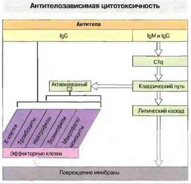
Fig. 24.1. All effector cells (K cells, platelets, neutrophils, eosinophils, and mononuclear phagocytes) bear Fc receptors through which they interact with antibodies bound to target cells. C3 activation can directly lead to complement-mediated lysis of target cells or promote the binding of phagocytes to their targets via C3b, C3bi, or C3d, which simultaneously activate the phagocytes.
Antibodies attached to The surface of cells or tissues can bind and activate the C1 component of complement, producing the following effects (see Chapter 4).
✵ Complement fragments (C3a and C5a) generated during activation recruit macrophages and polymorphonuclear cells to the site, and stimulate mast cells and basophils to release molecules that attract and activate other effector cells (see Chapter 10).
✵ Activation of the classical complement pathway and the Amplification loop lead to the deposition of C3b, C3bi, and C3d on the target cell membrane.
✵ Terminal activation of the classical complement pathway results in the assembly of the membrane attack complex (C5b-9), which inserts into the target cell membrane.
Effector cells—in this context, macrophages, neutrophils, eosinophils, and killer (K) cells—interact via their Fc receptors with cell-bound antibodies or via their C3 receptors with membrane-associated C3b, C3bi, and C3d (see Fig. 24.2 and Chapter 5). The binding of antibodies to phagocyte Fc receptors stimulates the production of increased amounts of Leukotrienes and Prostaglandins, which play a major role in the inflammatory process (see Chapter 5). Chemokines and chemotactic molecules, including C5a, leukotriene B4(LТВ4), and fibrin Peptides, can also activate cells newly arriving at the reaction site.

Fig. 24.2. Antibodies binding to membrane antigens on target cells opsonize them, thereby promoting phagocytosis. Cross-linking of antibodies with Fc receptors on the phagocyte surface activates the membrane oxidase complex of these cells, resulting in the release of oxygen radicals; simultaneously, protein phosphorylation is enhanced, leading to effector cell activation. Phospholipase A2 action releases arachidonic acid from membrane Phospholipids. Immune complexes induce the deposition of the complement fragment C3b, which can also interact with phagocyte receptors. Activation of the lytic cascade leads to the assembly of the membrane attack complex (MAC) from complement components C5-C9.
Effector cells that are firmly bound to target cells and fully activated can cause substantial tissue damage (see Chapter 10).
Antibodies of different isotypes vary in their ability to induce such reactions, depending on how efficiently they bind C1q or interact with effector cell Fc receptors. Complement fragments or IgG can act as opsonins by binding to host tissues or microorganisms; opsonized particles are then engulfed by phagocytes. By enhancing The activity of phagocytic lysosomal Enzymes and the production of highly reactive oxygen metabolites, opsonins increase the capacity of these cells not only to destroy pathogens but also to drive type II immunopathological hypersensitivity reactions (Fig. 24.3). For example, neutrophils derived from the synovial fluid of patients with rheumatoid Arthritis release higher amounts of superoxide anions upon stimulation compared to blood neutrophils. This is thought to result from the activation of neutrophils within inflamed joints by mediators such as immune complexes and complement fragments.

Fig. 24.3. Neutrophils are activated by cell-bound antibodies (IgG or IgA) and activated complement components. Each of these activators stimulates specific neutrophil functions. Note that activated C3 (including C3b, C3bi, and C3d—depending on the maturation state of the cells involved) and IgG mutually enhance each other's effects. Acting together, they generate an exceptionally potent activation signal for effector cells.
Effector cells cause the tissue injury characteristic of type II hypersensitivity against the body's own cells using the exact same mechanisms they employ against infectious agents (Fig. 24.4). For instance, most pathogenic microbes (unless resistant to phagocytic attack) are destroyed within phagolysosomes through the combined action of highly reactive oxygen and nitrogen metabolites, radicals, ions, altered pH, and other lytic factors. If the target is too large for phagocytosis, effector cells discharge the contents of their granules and Lysosomes toward the sensitized target (exocytosis) (Fig. 24.4). In certain cases, such as the eosinophil response to schistosome invasion (see Chapter 18), this granule release provides protection; however, when the target happens to be host cells sensitized by autoantibodies, this same reaction leads to damage of the host's own tissues (Fig. 24.5).

Fig. 24.4. The tissue-damaging effect of neutrophils on the host's own tissues is a reflection of the normal antibacterial function of these cells. 1. Neutrophils interact with microbes via their Fc and C3 receptors. 2. The microbial cell is then engulfed by the phagocyte, within which it is destroyed as lysosomes and the phagosome fuse to form a phagolysosome (3). In type II hypersensitivity reactions, individual antibody-coated host cells may also undergo phagocytosis; however, when the target is large, such as a basement membrane (I), neutrophils are unable to phagocytose it (II) and instead release their lysosomal contents extracellularly, damaging neighboring cells (III).
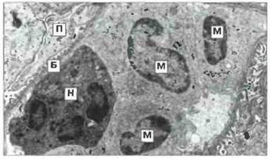
Fig. 24.5. Phagocytes attacking a basement membrane. The electron micrograph shows a neutrophil (N) and three monocytes (M) attached to the basement membrane (B) of a rabbit glomerular capillary in an animal with antibodies to the basement membrane. (P, podocyte.) ×3500. (Micrograph kindly provided by Prof. G.A. Andres.)
Antibodies also trigger hypersensitivity reactions by cross-linking K cells with target tissues. K cells are found primarily within the population of large granular lymphocytes and bind to antibodies via their high-affinity Fc receptors. In vitro, they exert cytotoxic effects on numerous cell types, mediated predominantly through cytokines and granule exocytosis. However, assessing THE CONTRIBUTION OF K cell activity to type II hypersensitivity reactions is rather difficult, partly because no two target cells possess equal susceptibility to a given effector cell. The susceptibility of target cells depends on the density of surface-expressed antigen and their capacity for damage repair. For instance, the lysis of a single erythrocyte requires only one membrane-attack complex, whereas the lysis of most nucleated cells demands many such complexes.
Below, we examine several scenarios in which type II hypersensitivity reactions are generally considered to be the primary cause of target cell destruction, i.e., immunopathology.
Last update: 13/08/2026
Editorial and Educational Adaptation: This material has been compiled based on the primary/original source text. The project team performed an editorial review, corrected technical inaccuracies, structured sections, and adapted the content for an educational format.
What was processed:
- elimination of formatting defects (OCR errors, structural breaks, corrupted characters);
- editorial organization of content;
- standardization of terminology in accordance with academic sources;
- verification of factual statements against the original source text.
All mentions of the author, publication year, and origin of the primary text have been preserved in accordance with the source.