Molecular Biology: Protein Structure and Functions - Stepanov V.M. 2005
Secondary Structure
Steric constraints and secondary structure of the peptide chain
The angles of rotation around the N—Ca and Ca—C(О) bonds are designated as φ and ψ, respectively (Fig. 5.2). The following convention is used to measure these dihedral angles. If one imagines an observer looking along the N—Ca bond, the "subsequent" Ca—C(О) bond may appear eclipsed by the "preceding" C(О)—N bond. Such a conformation (eclipsed or cisoid, which is inherently unfavorable sterically) corresponds to an angle of φ = 0°. If the Ca—C(О) bond is not eclipsed and the C(О)—N bond must be rotated clockwise to reach the eclipsed configuration, the angle φ is considered positive and increases up to 180°. In the latter case, a transoid configuration is reached. In the opposite direction, negative values of the angle φ are measured from 0° to -180°. Similarly, to determine the angle ψ, one looks along the Ca—C(О) bond, where the eclipsed conformation (in which the N—Ca bond obscures the C(О)—N bond) is assigned an angle of ψ = 0°, and the transoid configuration is assigned + or -180°.
The values of the φ and ψ angles for any given amino acid residue characterize its position within the Secondary Structure. Specifying these values for the entire Amino Acid Sequence determines how the polypeptide folds in three-dimensional space. As noted, any set of dihedral angle values for φ and ψ is theoretically possible; however, many of them are associated with significant steric hindrance and correspond to highly unfavorable energetic Conformations.
To describe the stereochemistry of amino acid residues in Proteins, the Ramachandran plot is a convenient tool. It is a square diagram where the values of the φ angle, ranging from -180° to +180°, are plotted on one axis, and the values of the ψ angle on the other (Figs. 5.3, 5.4). If no restrictions were imposed on the Stereochemistry of the polypeptide chain, the φ and ψ angles could assume any values, and the point representing their combination—and thus the conformation of a given amino acid residue—could fall anywhere on the map.
Class="center">
Fig. 5.2. Dihedral angles of the polypeptide chain.
The angle ψ is determined by the Rotation of the Ni—Cai bond around the Cai—C'i bond, viewed along the direction of the peptide chain, i.e., from Cai to the carbonyl carbon C'i, as indicated by the arrow. By convention, if the first bond obscures the C'i—Ni+1 bond (cisoid configuration), the angle is ψ = 0°; if these bonds are in the most distant positions (transoid configuration), the angle ψ is equal to +180° or -180°. Clockwise rotation is considered positive. The angle φ is determined by the rotation of the C'i-1—Ni bond around the Ni—Cai bond. The cisoid configuration, in which the bonds flanking Ni—Cai eclipse one another, corresponds to φ = 0°, and the transoid configuration corresponds to φ = ±180°. The figure depicts the so-called extended conformation of the peptide, in which both φ and ψ angles are equal to +180°.
In reality, not all combinations of φ and ψ angles are permissible. This is because any change in both angles alters the relative positions of atoms in neighboring amino acid residues. The spatial approach of atoms, especially bulky ones (oxygen, carbon, nitrogen), leads to their mutual repulsion. Overcoming this repulsion requires significant Energy Expenditure, rendering such a polypeptide chain configuration unstable. Ramachandran plots are analogous to geographical maps, featuring contour lines corresponding to specific energy levels. More favorable conformations correspond to lower energy levels, which appear as valleys or depressions on the Ramachandran plot, whereas the least favorable ones correspond to ridges.
Let us examine the Ramachandran plot for Glycine residues, which lack a side chain (see Fig. 5.3). In this case, a fairly large portion of the map is allowed, permitting a wide range of φ and ψ angles. However, conformations where these angles are close to zero are unfavorable: this corresponds to the eclipsed configuration, where the steric clash of carbonyl oxygen atoms is particularly pronounced. The Ramachandran conformational plot for glycine is simple—it reveals a cross-shaped "forbidden" elevation along the axes where φ = 0° and ψ = 0°, alongside quite extensive depressions covering at least half of the total surface area.
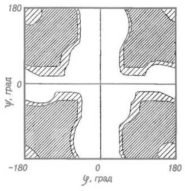
Fig. 5.3. Ramachandran plot for glycine.
The potential energy is plotted for a pair of peptide units with a glycine residue between them (isoeenergetic lines). For glycine, conformations occupying the cross-shaped elevation on this map near φ and ψ values of 0° are unfavorable, as this corresponds to the eclipsed configuration; extensive depressions (shaded) represent allowed regions. Compare Fig. 5.13, which shows that glycine residues in real proteins allow adjacent peptide bonds to adopt vastly different conformations relative to one another.
This property of glycine as the residue with the fewest conformational restrictions plays a specific role in forming the Spatial Structure of Proteins. Characteristically, on Ramachandran Plots for real proteins, the points corresponding to glycine residues do not cluster into two main valleys, as is typical for Other Amino Acids, but are distributed across almost the entire field. Obviously, glycine residues are utilized at points in the protein's spatial structure where a non-standard conformation is required, particularly at sharp turns of the polypeptide chain. It is no coincidence that glycine residues, which might seem incapable of directly participating in protein function due to the lack of a side chain, more often than not remain invariant across families of evolutionarily related proteins, highlighting their indispensability in structure formation.
Obviously, the set of allowed conformations decreases significantly if one moves away from the simplified model considered thus far and accounts for the steric effects of amino acid side chains. Analyzing THE CONTRIBUTION OF all 19 side chains would be challenging, but it is unnecessary. The most significant constraints on the conformational freedom of amino acid residues are imposed by the simplest substituent at the Ca atom—the methyl group in the Alanine residue. Further complexities of the side chain occur relatively far from the main chain and have little influence on its Selection of an optimal conformation. The Ramachandran plot for alanine serves as a sufficiently accurate approximation for other Amino acids as well.
This map (Fig. 5.4) reveals two valleys corresponding to stable conformations—i.e., optimal combinations of φ and ψ angles—and a shallow depression. One of the valleys corresponds to the right-handed a-helix characteristic of proteins (φ = -57°, ψ = -47°), while the other corresponds to the parallel ß-Structure (φ = -119°, ψ = +113°) and the antiparallel ß-structure (φ = -139°, ψ = +135°).

Fig. 5.4. Ramachandran plot for L-alanine.
The potential energy distribution for an alanine residue flanked by two peptide units is shown in φ and ψ coordinates (see caption to Fig. 5.3). The deep valley in the upper left quadrant corresponds to the conformation characteristic of the ß-structure, whereas the valley connected to it by a col in the lower left quadrant corresponds to the right-handed a-helix (aR) commonly found in proteins. A minor depression in the upper right quadrant corresponds to the extremely rare left-handed a-helix (aL). It should be noted that the plot can be toroidally wrapped around the φ, ψ = 0° axes; therefore, for instance, the valley corresponding to the ß-structure continues at the lower left edge of the map.
The fact that ordinary amino acids allow for only two regions—two valleys—on the conformational map, and consequently only two sets (within certain tolerances) of φ and ψ angles, largely dictates the limited repertoire of periodic secondary structures. Thus, by attaching another amino acid residue with a similar set of dihedral angles (φ = -57°, ψ = -47°) to a first one, followed by a third and so on, one obtains a periodic secondary structure—the a-helix. Similarly, linking residues with two other allowed sets of angles leads to the two known types of ß-structures.
Last update: 13/08/2026
Editorial and Educational Adaptation: This material has been compiled based on the primary/original source text. The project team performed an editorial review, corrected technical inaccuracies, structured sections, and adapted the content for an educational format.
What was processed:
- elimination of formatting defects (OCR errors, structural breaks, corrupted characters);
- editorial organization of content;
- standardization of terminology in accordance with academic sources;
- verification of factual statements against the original source text.
All mentions of the author, publication year, and origin of the primary text have been preserved in accordance with the source.