Principles of Protein Structural Organization - H. Schultz 1982
Models, Depiction, and Documentation of Protein Structures
Abstract Concepts of Chain Folding
Ramachandran Plots for Main-Chain Angles
Up to this point, we have discussed representing molecular structures using the Cartesian coordinates of their constituent atoms. However, for certain Applications—particularly in the theoretical Analysis of Protein structures—it is necessary to employ other, more Abstract representations.
The polypeptide backbone is fully described by a set of (φ, ψ) angles. As discussed in Chapter 2, chain folding can be completely characterized by the backbone dihedral angles φ and ψ at each Ca-atom (ψ only for the N-terminal Ca-atom, and φ only for the C-terminal Ca-atom). These angles can be plotted as points on a (φ, ψ) map (Fig. 2.4); however, to identify the angles, all points must be labeled with the sequence numbers of the corresponding Ca-atoms. Such a map becomes cumbersome and difficult to read, making it an unsatisfactory representation of the Structure.
Class="center">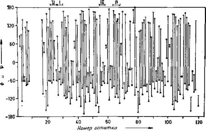
Fig. 7.9. Linear representation of the backbone dihedral angles for the N-terminal region of Chymotrypsin [402].
A black dot corresponds to the value of φ, and an open circle to ψ; The values of both angles for each residue are connected by a line. Secondary structures are clearly discernible. The figure highlights type I, II, and III reverse turns (beta-turns) as well as the β-structure. α-Helices appear as consecutive short lines near —60°.
A more convenient approach was proposed by Balasubramanian [402]. Based on the (φ, ψ) map, he derived a linear plot showing the dependence of the φ and ψ angles on the residue sequence number, as illustrated in Fig. 7.9. This plot has the advantage of allowing the (φ, ψ) angles of any given residue to be determined with ease. Furthermore, it effectively highlights regions of Secondary structure.
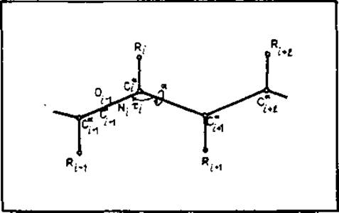
Fig. 7.10. Representation of the polypeptide backbone using virtual bonds between Ca atoms. The chain is completely described by the virtual dihedral angles αi, defined by the Cai-1, Cai, Cai+1 atoms, and the virtual valence angles τi. This description was used to construct a wireframe model of the backbone (Fig. 7.3, c). In a simplified approximation, τi can be considered as a function of αi [30], in which case the backbone is described by only a single independent parameter per residue.
The Ca—Ca virtual bonds simplify the representation of chain folding. In an alternative approach, the peptide backbone is represented solely by Ca-atoms and virtual bonds connecting them, as shown in Fig. 7.10. Chain conformation in this case is described by virtual valence and dihedral angles [30, 403]. Peptide bond geometry restricts the virtual valence angle to a range of approximately 80° to 160°, whereas the virtual dihedral angle is initially unconstrained. However, due to steric clashes, only about half of the theoretically possible angle combinations are actually realized. This representation has been used to model chain folding [404] and is also useful for constructing wireframe models (Fig. 7.3, c). A more detailed Description of the backbone using virtual Ca bonds is discussed in [403] and [405].
Last update: 06/08/2026
Editorial and Educational Adaptation: This material has been compiled based on the primary/original source text. The project team performed an editorial review, corrected technical inaccuracies, structured sections, and adapted the content for an educational format.
What was processed:
- elimination of formatting defects (OCR errors, structural breaks, corrupted characters);
- editorial organization of content;
- standardization of terminology in accordance with academic sources;
- verification of factual statements against the original source text.
All mentions of the author, publication year, and origin of the primary text have been preserved in accordance with the source.