IMMUNOLOGY TEXTBOOK - Mercury Podillya 2013
ACQUIRED IMMUNODEFICIENCY STATES
Secondary immunodeficiencies in chronic inflammatory processes of the bronchopulmonary system
In patients with Chronic Bronchitis and various immune status alterations, remission was achieved through Treatment with polyoxidonium or lycopid. Polyoxidonium is best prescribed during the acute phase in combination with antibacterial therapy when abnormalities in the lymphoid system and phagocytosis are observed. Positive results in treating infection flare-ups in Bronchial Asthma patients have been achieved using these immunomodulators.
As an example of T-Cell link and phagocytosis impairments in bronchopulmonary pathology, we present the clinical case of patient S., aged 52, who underwent treatment in the therapeutic department with a Diagnosis of chronic bronchitis, acute exacerbation; immune system dysfunction with predominant impairments of the T-cell link and phagocytosis.
Patient S., aged 52, complains of a cough with a small amount of mucopurulent sputum and mixed dyspnea upon moderate physical exertion. He has been ill for 5 years, with the latest exacerbation lasting 1 week. Physical examination reveals slight diffuse cyanosis. Percussion reveals a box-like Resonance note in the lower lung fields; Auscultation reveals dry, scattered rales over the entire lung surface against a Background of harsh breathing.
Sputum analysis: volume 15 ml, mucopurulent, without foreign impurities; leukocytes – 30-40 per high-power field (HPF), neutrophils 20-30 per HPF, macrophages – 8-10 per HPF, erythrocytes – 1-2 per HPF, epithelial Cells (columnar) – 1-2 per HPF; mixed flora – St. pneumoniae, H. influenzae. AFB (acid-fast bacilli) not detected.
Conclusion of sputum analysis: mucopurulent sputum with an elevated neutrophil count and the presence of Bacteria. Spirometry findings (Fig. 8, Table 73).
Class="center">Table 73. Spirometry
Parameter |
Actual |
Predicted |
% |
Comment |
VC (L) |
2.28 |
5.13 |
42 |
Extremely sharp decrease |
FVC (L) |
2.13 |
4.97 |
42 |
Extremely sharp decrease |
FEV1 (L) |
2.11 |
4.15 |
52 |
Significant decrease |
FEV1/VC |
92,51 |
81,88 |
116 |
Slight increase |
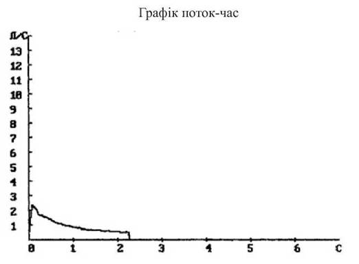
Fig. 8. Spirometry conclusion: pronounced impairment of pulmonary ventilatory function, predominantly of a restrictive type, with a sharp decrease in vital capacity
Chest X-ray (straight projection): enhanced bronchovascular markings, thickened lung roots, and emphysematous changes in both Lungs.
Chest X-ray conclusion: signs of chronic bronchitis.
Immunogram of patient S., aged 52: neutrophilic leukocytosis with a moderate left shift of the differential. Relative lymphocytopenia due to a decrease in CD4+ T-helper cells and CD8+ cytotoxic T-lymphocytes; immunoregulatory index (IRI) 1.25. Elevated levels of B-lymphocytes and production of IgM, IgG, and IgA IMMUNOGLOBULINS. NK cells are within normal limits. A slight increase in the phagocytic capacity of neutrophils (phagocytic index, phagocytic number) along with a sharp decrease in the functional reserve of the oxidation-reduction potential of phagocytes (NBT test res. < 16), indicating incomplete phagocytosis (Table 74).
Diagnosis: chronic bronchitis, acute exacerbation. Diffuse pulmonary sclerosis. Pulmonary emphysema. Respiratory failure grade II. Immune system dysfunction with predominant impairments of the T-cell link and phagocytosis.
Conclusion: T-cell dysfunction combined with ineffective phagocytosis. Signs of an acute chronic inflammatory process (elevated IgG, IgM) at the mucous membrane level (increased IgA).
Based on the immunological status features of patient S., the following immunotropic therapy regimen was prescribed for the treatment of chronic bronchitis:
1) etiotropic antibacterial therapy – amoxiclav 500/125 mg 3 times a day IV, 7 days;
2) galavit 0.1 g IM every other day, 10 injections;
3) immunofan 0.005%, 0.4 – 0.7 ml SC twice a week, 10 injections;
4) ACC-long 600 mg, 1 tab. once daily, 10 days.
Immunorehabilitation:
5) respibron 1 tab. daily sublingually, course – 10 days. For Prevention, 1 tab. daily, course – 10 days for 3 months;
6) thymalin 1 ml subcutaneously every other day, 10 days.
Table 74. Immunogram of patient S., aged 52
Parameter |
Result |
Reference Range |
||||||
142 |
F - 115 - 145, M - 132 - 164 g/L |
Pronounced anisocytosis, anisochromia TSI=45% |
||||||
Erythrocytes |
5.1 |
F - 3.7 - 4.7, M - 4.0 - 5.1×10¹²/L |
||||||
Platelets |
250 |
150 - 320×109/L |
||||||
ESR |
25 |
2 - 15 mm/h |
||||||
Leukocytes |
10.3 |
4 - 9×10⁹/L |
||||||
Neutrophils |
Band |
Segmented |
Eosinophils |
Basophils |
Monocytes |
Lymphocytes |
LAL |
Plasma |
43 - 71 % |
1 - 4 % |
0.5 - 5% |
0 - 1% |
3 - 9% |
25 - 37% |
1-5% |
0 - 1% |
|
2000-6500 |
80-400 |
80-370 |
20-80 |
90-720 |
1600-3000 |
80-500 |
20-80 |
|
76 |
10 |
66 |
4 |
1 |
3 |
16 |
0 |
0 |
7830 |
100 |
7730 |
410 |
100 |
300 |
1648 |
||
Immunological parameters |
Result |
Norm |
Immunological parameters |
Result |
Norm |
|||
(SI units) |
(SI units) |
|||||||
T-Lymph. |
% |
40 |
50 - 80 |
Ig G |
18.5 |
8.0-18.0 |
||
CD-3 |
Absolute count |
656 |
1000-2200 |
g/L |
||||
T-helper |
% |
20 |
33-46 |
Ig M |
2.2 |
0.2-2.0 g/L |
||
CD-4 |
Absolute count |
328 |
309-1571 |
|||||
T-suppressor |
% |
16 |
17-30 |
Ig A |
4.0 |
0.3-3.0 g/L |
||
CD-8 |
Absolute count |
262 |
282-999 |
|||||
IRI |
CD-4/CD-8 |
1.25 |
1.4-2.0 |
CIC |
90 |
30 - 50 units |
||
opt. density |
||||||||
NK cells CD-16 |
% |
20 |
12 - 23 |
Engulfing |
PNI |
85 |
60 - 80% |
|
Absolute count |
328 |
72-543 |
activity |
PI |
3.82 |
1.5 - 3.5 |
||
B-lymph. |
% |
32 |
17-31 |
NBT test |
spon. |
5 |
up to 10% |
|
CD-22 |
Absolute count |
525 |
109-532 |
ind. |
9 |
- |
||
LST |
spon. |
9 |
up to 10% |
res. |
4 |
h16% |
||
ind. |
45 |
50-70% |
CH-50 |
45 |
30 - 60 |
|||
hem. units/mL |
||||||||
In immunodeficiencies manifesting against the background of COPD, a positive effect is observed with the administration of levamisole, T-activin, sodium nucleinate, diucifon, and others. In A number of cases in COPD, the inhalation route of immunomodulator administration is preferable (a combination of dimexide and levamisole solutions).
As an example of a B-cell type immunodeficiency in bronchopulmonary pathology, we present the case history of patient T., aged 58, who was treated in the therapeutic department with a diagnosis of: chronic obstructive pulmonary disease, stage II, exacerbation, stage II respiratory failure.
Patient T., aged 58 (Table 75), has been suffering from COPD for the past 10 years, smokes up to 10 cigarettes a day despite warnings, and experiences disease exacerbations 2 to 3 times a year. The patient is continuously maintained on basic therapy with Spiriva (tiotropium bromide) 18 mcg, 1 inhalation once daily, and theophylline 150 mg once daily.
Immunogram of patient T., aged 58: relative lymphocytopenia. Dysregulation of the T-cell link with a relative increase in the T-helper population, IRI 2.5. Decreased B-lymphocyte level. IgM production is decreased, while IgA is increased. NK cells are within normal limits. Significant increase in the engulfing capacity of neutrophils (PI, PNI) alongside a decrease in the functional reserve of the phagocyte oxidation-reduction potential (NBT test res. < 16), indicating incomplete phagocytosis.
Diagnosis: chronic obstructive pulmonary disease, stage II, exacerbation. Diffuse pneumosclerosis. Pulmonary emphysema. Stage II respiratory failure. B-cell type immunodeficiency (D 84.9).
Conclusion: immunodeficiency state of the B-cell link, dysregulation of immunoglobulin production, indicating an exacerbation of the chronic inflammatory process (elevated IgM) in the mucosal area (increased IgA). Activation of phagocytosis with signs of its inefficiency.
Final diagnosis: chronic obstructive pulmonary disease, stage II, exacerbation. Diffuse pneumosclerosis. Pulmonary emphysema. Stage II respiratory failure. B-cell type immunodeficiency (D 84.9).
Based on the immunological status features of patient T., the following immunotropic therapy regimen was prescribed for the treatment of COPD:
1) etiotropic antibacterial therapy - levofloxacin 500 mg IV drip once daily for 7 days, dimexide 5 mL in 200 mL of 0.9% sodium chloride solution IV drip once daily for 5 days; azithromycin 500 mg once daily for 3 days;
2) polyoxidonium 6 mg IM twice a week for 10 days;
3) galavit 100 mg once daily IM for 10 days;
4) lactofiltrum 2 caps. twice daily for 14 days.
5) fluconazole 100 mg every other day for 10 days.
Immunorehabilitation:
6) IRS-19 inhalations once daily for 20 days;
7) thymalin 1 mL subcutaneously every other day for 10 days.
Table 75. Immunogram of patient T., aged 58
Parameter |
Result |
Reference Range |
||||||
Hemoglobin |
122 |
F - 115 - 145, M - 132 - 164 g/L |
Pronounced anisocytosis, anisochromia TSI=45% |
|||||
Erythrocytes |
3.8 |
F - 3.7 - 4.7, M - 4.0 - 5.1×10¹²/L |
||||||
Platelets |
220 |
150 - 320×109/L |
||||||
ESR |
11 |
2 - 15 mm/h |
||||||
Leukocytes |
6.6 |
4 - 9×10⁹/L |
||||||
Neutrophils |
Band |
Segmented |
Eosinophils |
Basophils |
Monocytes |
Lymphocytes |
LAL |
Plasma |
43 - 71 % |
1 - 4 % |
0.5 - 5% |
0 - 1% |
3 - 9% |
25 - 37% |
1-5% |
0 - 1% |
|
2000-6500 |
80-400 |
80-370 |
20-80 |
90-720 |
1600-3000 |
80-500 |
20-80 |
|
66 |
3 |
63 |
6 |
1 |
3 |
23 |
0 |
0 |
4360 |
200 |
4160 |
400 |
70 |
200 |
1518 |
||
Immunological parameters |
Result |
Norm |
Immunological parameters |
Result |
Norm |
|||
(SI units) |
(SI units) |
|||||||
T-lymph. |
% |
72 |
50 - 80 |
Ig G |
11.9 |
8.0-18.0 |
||
CD-3 |
Absolute count |
1092 |
1000-2200 |
g/L |
||||
T-helper |
% |
53 |
33-46 |
Ig M |
0.18 |
0.2-2.0 g/L |
||
CD-4 |
Absolute count |
804 |
309-1571 |
|||||
T-suppressor |
% |
21 |
17-30 |
Ig A |
4.8 |
0.3-3.0 g/L |
||
CD-8 |
Absolute count |
319 |
282-999 |
|||||
IRI |
CD-4/CD-8 |
2.5 |
1.4-2.0 |
CIC |
59 |
30 - 50 units |
||
opt. density |
||||||||
NK cells CD-16 |
% |
23 |
12 - 23 |
Engulfing |
PNI |
91 |
60 - 80% |
|
Absolute count |
349 |
72-543 |
activity |
PI |
4.45 |
1.5 - 3.5 |
||
B-lymph. |
% |
8 |
17-31 |
NBT test |
spon. |
21 |
up to 10% |
|
CD-22 |
Absolute count |
121 |
109-532 |
ind. |
26 |
- |
||
LST |
spon. |
10 |
up to 10% |
res. |
5 |
h16% |
||
ind. |
40 |
50-70% |
Complement |
CH-50 |
55 |
30 - 60 |
||
hem. units/mL |
||||||||
Last update: 13/08/2026
Editorial and Educational Adaptation: This material has been compiled based on the primary/original source text. The project team performed an editorial review, corrected technical inaccuracies, structured sections, and adapted the content for an educational format.
What was processed:
- elimination of formatting defects (OCR errors, structural breaks, corrupted characters);
- editorial organization of content;
- standardization of terminology in accordance with academic sources;
- verification of factual statements against the original source text.
All mentions of the author, publication year, and origin of the primary text have been preserved in accordance with the source.