GENERAL MICROBIOLOGY - T.P. Pyrog - 2004
8. FUNGI
8.4. REPRODUCTION IN FUNGI
The reproductive Organs of Fungi are spores. Spores can be motile (planospores, zoospores) and non-motile (aplanospores).
Fungi are characterized by the following modes of reproduction: vegetative, asexual, and sexual.
Vegetative Reproduction. This occurs through hyphal fragmentation, budding, and The formation of chlamydospores (which are capable of germination).
Asexual reproduction. Organs of asexual reproduction (spores) can be endogenous or exogenous. Endogenous spores develop inside specialized structures (sporangia) and are called spores (sporangiospores). Exogenous spores are produced at the tips of differentiated hyphal branches (conidiophores) and are called conidia (conidiospores) (see Chapter 3, Fig. 3.4).
Conidial sporulation (reproduction via conidia) is predominantly characteristic of deuteromycetes.
Sexual reproduction. Sexual reproduction in fungi, as in other eukaryotes, involves the fusion of two nuclei. The process of sexual reproduction can be divided into three phases. First, plasmogamy takes place—the merging of two protoplasts. The resulting Cell contains two nuclei. This pair of nuclei (a dikaryon) does not necessarily fuse immediately. During subsequent divisions, Cells may remain in the dikaryotic phase. In this process, the two nuclei divide simultaneously (conjugate division). Later on, often only after the formation of a fruiting body, the two haploid nuclei fuse (the second phase of sexual reproduction, karyogamy), forming a diploid zygote Nucleus. The third phase is Meiosis, or reduction division, which restores the original chromosome number (haploid). These three phases (plasmogamy, karyogamy, and meiosis) occur consecutively in some fungi, while in others they take place at different Stages of the fungal life cycle.
The organs of sexual reproduction in fungi are spores (oospores in oomycetes, zygospores in zygomycetes, ascospores in ascomycetes, and basidiospores in Basidiomycetes).
The sexual reproduction phase begins with the formation of Gametes, which are produced within specialized, morphologically differentiated cells called gametangia. If the gametes originating from the male and female parental cells are morphologically indistinguishable, they are called isogametes. Isogametes are characteristic of zygomycetes. In oomycetes, the gametes are morphologically distinct: the male gametangia (and corresponding gametes) are called antheridia, while the female ones are called oogonia. If male and female gametangia are produced on the same vegetative body developed from a single spore, the fungus is referred to as homothallic (hermaphroditic). In heterothallic fungi, the thalli are sexually differentiated, bearing either exclusively male or exclusively FEMALE REPRODUCTIVE ORGANS.
Oospores are formed through the fusion of a morphologically distinct female gamete from an oogonium and a male gamete from an antheridium (a process known as heterogamy), whereas zygospores are formed by morphologically identical female and male gametes (known as isogamy).
A distinctive feature of the Sexual process in lower fungi is that Fertilization yields a zygote, which Functions as a resting spore. Reduction division (meiosis) occurs exclusively during the germination of such a spore, restoring the haploid set of Chromosomes in The Nucleus.
Fig. 8.2 illustrates asexual (via conidia) and sexual reproduction in higher fungi, specifically ascomycetes. During sexual reproduction, gametangia 2 develop on the mycelium, consisting of an antheridium (containing "+" nuclei, the male gamete) and an ascogonium (containing "-" nuclei, the female gamete). Within the ascogonium, the nuclei pair up but do not fuse. The fertilized ascogonium gives rise to dikaryotic ascogenous hyphae 3, and the pairs of nuclei undergo mitosis, replicating new paired chromosomes. Subsequently, karyogamy takes place at the tips of the ascogenous hyphae 4, after which the diploid nuclei undergo meiosis. This results in eight haploid nuclei that develop into ascospores enclosed within an ascus 4. Simultaneously, asci incorporated into the mycelial hyphae transform into an ascocarp 5. The ascocarp disintegrates into individual asci containing eight ascospores each. The ascospores germinate to form a new mycelium.
Class="center">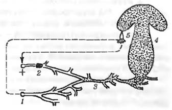
Fig. 8.2. Reproduction of Ascomycetes
The sexual process in basidiomycetes (Fig. 8.3) is characterized by the initial formation of a dikaryon (which can occur via the fusion of two haploid vegetative cells or through nuclear division within a single cell). Subsequently, the cells containing the dikaryon develop into a complex dikaryophytic mycelium, which gives rise to fruiting bodies. Basidia develop either on the fruiting bodies or directly from the mycelium. Concurrently with their formation, karyogamy of the dikaryon occurs (nuclear fusion followed by reduction division, most commonly into four nuclei), initiating the formation of four basidia, which in turn produce basidiospores. Germinating basidiospores give rise to a haploid mycelium composed of mononuclear cells, which eventually re-establish the dikaryon and the dikaryophytic mycelial phase (see Fig. 8.3).
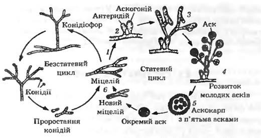
Fig. 8.3. Developmental cycle of a basidiomycetous fungus:
1 — basidiospores; 2 — haploid mycelium; 3 — dikaryophytic mycelium; 4 — fruiting body originating from the dikaryophytic mycelium; 5 — basidia with basidiospores
Thus, a defining feature of the sexual process in basidiomycetes is the presence of a dikaryophytic mycelial phase, which represents a distinct variation from the typical diploid mycelium. Furthermore, basidiospores are produced exogenously on sterigmata (outgrowths) of the basidium.
Last update: 13/08/2026
Editorial and Educational Adaptation: This material has been compiled based on the primary/original source text. The project team performed an editorial review, corrected technical inaccuracies, structured sections, and adapted the content for an educational format.
What was processed:
- elimination of formatting defects (OCR errors, structural breaks, corrupted characters);
- editorial organization of content;
- standardization of terminology in accordance with academic sources;
- verification of factual statements against the original source text.
All mentions of the author, publication year, and origin of the primary text have been preserved in accordance with the source.