GENERAL MICROBIOLOGY - T.P. Pyrog - 2004
3. MORPHOLOGY OF MICROORGANISMS
3.1. BACTERIA
Based on their shape, these organisms are divided into several groups: spherical, cylindrical, spiral, unusual shapes, and filamentous.
Spherical Bacteria, or cocci (from Greek kokkos – grain), have a rounded shape. Depending on the arrangement of Cells after division, they are grouped into various categories (Fig. 3.1).
Micrococci (Micrococcus) (from Greek mikros – small) are cocci that divide in a single plane and, after division, remain solitary; for example, Micrococcus aqua is a common aquatic inhabitant (Micrococcus is the genus name, and aqua is the species epithet).
Diplococci (Diplococcus) (from Greek diploos – double) are cocci that divide in a single plane and remain in pairs after division; Examples include Methylococcus capsulatus, a methane-oxidizing bacterium, and Neisseria gonorrhoeae, the CAUSATIVE AGENT OF Gonorrhea.
Streptococci (Streptococcus) (from Greek streptos – twisted, chain) are cocci that divide in a single plane. After division, a connection remains between the cells, arranging them into chains, such as Streptococcus lactis, a lactic acid bacterium that causes milk to sour. The chains can be short (3–4 cells) or long (several dozen cells).
Class="center">
Fig. 3.1. Shapes of bacterial cells:
1 — micrococcus; 2 — diplococcus; 3 — streptococcus; 4 — tetracoccus; 5 — sarcina; 6 — staphylococcus; 7 — small rod; 8 — large rods connected in a chain; 9 — ovoid cocci; 10 — trichome cells; 11 — vibrio; 12 — spirochete; 13 — worm-like Cell; 14 — multicellular form; 15 — star-shaped; 16 — rods positioned at an angle to each other; 17 — triangular; 18 — branching forms; 19 — toroidal; 20 — budding cells; 21 — dumbbell-shaped; 22 — non-cylindrical trichomes; 23 — tapered rods; 24 — bean-shaped diplococcus; 25 — stalked cell; 26 — plate-like archaeal cells; 27 — spirillum; 28 — amoeboid; 29 — lanceolate diplococcus; 30 — cell with hyphae forming buds; 31 — loop-forming chain of cells; 32 — cells joined into plates; 33 — star-shaped rosette of cells; 34 — tuberoid cells; 35 — bacterial mucilaginous stalks; 36 — spined cell
Tetracocci (Tetracoccus) (from Greek tetra – four) are cocci that divide in two mutually perpendicular planes, forming tetrads after division. An example is Aerococcus.
Sarcinae (Sarcina) (from Latin sarcio – to bind/patch) are cocci that divide in three mutually perpendicular planes and, following division, form packets of 8, 16, 32, or 64 cells. Examples include Sarcina flava (yellow sarcina) and Methanosarcina methanica (methane-producing sarcina).
Staphylococci (Staphylococcus) (from Greek staphyle – bunch of grapes) are cocci that divide in multiple planes and form grape-like clusters after division. For instance, Staphylococcus aureus is the causative agent of purulent infections.
Cocci are not always perfectly round; they can be lanceolate (Streptococcus pneumoniae, the causative agent of Pneumonia), oval (Peptostreptococcus), or elongated (Ruminococcus).
Most cocci are non-motile and do not form endospores, although some may possess flagella (Planococcus, Planosarcina) or endospores (Sporosarcina).
The cylindrical bacterial shape (from Greek bacteria, Latin bacillum – rod) is characteristic of the majority of bacteria. Rod-shaped bacteria are divided into endospore-forming (Bacillus, Clostridium) and non-endospore-forming taxa (Pseudomonas, Xanthomonas). Rod forms vary in length, transverse diameter, end Morphology, and cellular arrangement. They can be long (exceeding 3 µm) (Bacillus megaterium, a putrefactive bacterium; Clostridium botulinum, the causative agent of botulism), short (1 µm) (enteric group bacteria), or very short (less than 1 µm), where the length barely exceeds The Cell diameter, leading to their designation as coccobacilli (Brucella abortus, the causative agent of brucellosis). Based on transverse diameter, bacteria are categorized as thin (Mycobacterium tuberculosis, the causative agent of tuberculosis) and thick (Escherichia coli). The ends of the rods may be rounded, truncated, tapered, or thickened. Rods are arranged singly, in pairs (Pseudomonas), or in chains (Bacillus mycoides, a typical soil bacterium). Some rods lie at an angle to one another, forming X- or Y-like figures, such as arthrobacteria, corynebacteria, nocardiae, and mycobacteria.
Spiral-shaped bacteria differ in the number and nature of their turns, as well as cell length and thickness. They can be divided into rigid forms (vibrios, spirilla) and flexible, undulating forms (spirochetes).
Vibrios (from French vibrion) resemble a curved rod or comma (Vibrio cholerae, the causative agent of cholera).
Spirilla (from Latin spira – coil) are spirally curved cells with a large transverse diameter and a small number of high coils (Spirillum volutans, a saprophytic bacterium; Spirillum minus, a pathogenic species capable of causing disease).
Spirochetes (from Latin spira – coil, Greek chaite – Hair) are flexible, slender, spirally curved cells (Fig. 3.2).
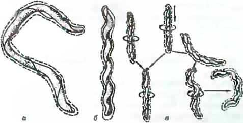
Fig. 3.2. Diagram of the movement of a flexible spirochete cell: a — axial filament; b, c — contraction of the axial filament
They consist of an outer sheath, a protoplasmic cylinder, and axial filaments. The axial filaments wind around the protoplasmic cylinder and are intracellular structures located within the periplasmic space. The axial (or periplasmic) filaments of spirochetes are functional analogs of bacterial flagella. Examples include Spirochaeta plicatilis, a common inhabitant of fresh, marine, and sewage waters; Treponema pallidum, the causative agent of Syphilis; and Treponema macrodentium, an oral spirochete found in dental caries.
Bacteria with unusual shapes exhibit great morphological diversity. Toroidal (closed or open rings), star-shaped, and tuberoid cells are illustrated in Fig. 3.1. The geometry of flat square plates and box-like flat cells of diverse shapes is characteristic of archaebacteria.
Filamentous forms of bacteria (trichomes, from the Greek trichoma meaning hair) are mostly rod-shaped cells joined into long chains united by mucus, sheaths, plasmodesmata (bridges), or a common envelope. Typically, the exterior of a trichome is covered with additional envelopes that do not participate in forming partitions between cells. The cells of trichomes are predominantly rod-shaped.
All bacteria are characterized by a constant cell shape due to the Structural Features of one of their envelopes — The Cell wall. However, some bacteria exhibit polymorphism. These include Mycoplasmas and L-forms, whose cells lack a cell wall, as well as arthrobacter, nocardioform bacteria, and corynebacteria, which undergo a morphological cycle during their development: coccus — rod — coccus.
Bacterial cell sizes vary significantly. The diameter of spherical bacteria ranges from 0.2 to 2.5 µm. The smallest are mycoplasmas at 0.15 µm. This size represents the theoretical lower limit for the Cellular Organization of life, at which a cell can still contain the minimum number of protein molecules (about 1,200) and the minimum set of enzymatic reactions required to maintain cellular Structure. Rod-haired bacteria have a thickness of 0.5–1.0 µm and a length ranging from 1–2 to 10 µm. Filamentous forms can reach macroscopic sizes (1 mm) and are visible to the naked eye. The length of spirochetes ranges from 1–3 to 100–500 µm. The lower size limit of unicellular bacteria is determined by the space required to pack the machinery ensuring the cell's independent existence, while the upper limit is governed by the optimal surface-area-to-volume ratio.
Last update: 12/08/2026
Editorial and Educational Adaptation: This material has been compiled based on the primary/original source text. The project team performed an editorial review, corrected technical inaccuracies, structured sections, and adapted the content for an educational format.
What was processed:
- elimination of formatting defects (OCR errors, structural breaks, corrupted characters);
- editorial organization of content;
- standardization of terminology in accordance with academic sources;
- verification of factual statements against the original source text.
All mentions of the author, publication year, and origin of the primary text have been preserved in accordance with the source.