Medical Genetics - V. M. Zaporozhan 2005
Etiology of Hereditary Diseases
Non-Mendelian Inheritance in Humans
Sex Chromatin (Barr Bodies)
Random inactivation of one of the X Chromosomes during embryonic development (X-Chromatin). Sex chromatin (also known as X-chromatin or Barr body) is a condensed X-chromosome discovered in 1949 by Barr and Bertram. The condensed X chromosome appears as a dense, well-staining oval body measuring 0.8–1.1 µm. It is typically located at the periphery of the interphase Nucleus in female mammals and is absent in males (Fig. 2.16). In buccal epithelial Cells, the Barr body lies just beneath the nuclear envelope. In neutrophil leukocytes, it takes the shape of a drumstick. The theory regarding THE ORIGIN OF Barr bodies (condensed X chromosomes) was proposed by the English geneticist Mary Lyon.
The formation of sex chromatin is a mechanism of Gene dosage regulation in all mammals, particularly in humans. Normally, females have two X chromosomes, whereas males have one. The length of the X chromosome is 6.8 µm, and that of the Y chromosome is 2.8 µm. Consequently, females possess more genes than males. Normal Human Development requires a specific gene dosage. Therefore, evolution has shaped a robust, specialized mechanism to compensate for differences in the dosage of X-linked genes. The major part of one of the X chromosomes in female embryos condenses and is inactivated on the 16th day of embryonic development, while the remainder remains active. X-chromosome inactivation occurs simultaneously in all embryonic cells, except for germ Cell precursors, where both X chromosomes remain active. Inactivation is associated with gene methylation. In each individual cell, this process is entirely random: in one cell of a female embryo, the maternal X chromosome is inactivated, whereas in another, the paternal one is. All descendants of these cells will carry the same inactivated chromosome.
This results in a unique phenomenon of functional mosaicism in the female body, which essentially consists of two cell clones. In the cells of one clone, the maternal X chromosome is active, while in the Cells of the other, the paternal one Functions. If both X chromosomes carry normal genes, this mosaicism is undetectable. However, if a woman is a heterozygous carrier of an X-linked mutant gene, a normal gene may be expressed in one part of her cells, and a pathological one in another. For instance, in ocular albinism—an X-linked recessive disorder—affected males lack retinal pigment, and the fundus appears pale. Heterozygous females exhibit irregular retinal pigmentation, with pigmented and depigmented patches visible on the fundus. Another example of an X-linked recessive disorder is anhidrotic ectodermal Dysplasia. Affected males lack Sweat Glands and present with hypotrichosis and oligodontia. Heterozygous females display both normal Skin areas and patches devoid of sweat glands.
Class="center">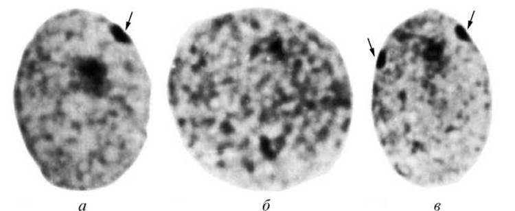
Fig. 2.16. Sex chromatin in epithelial cell nuclei: a — a single Barr body in The Nucleus of a healthy female; b — nucleus of a healthy male; c — two Barr bodies in the nucleus of a female with a 47,XXX karyotype
Clinical significance of inactivation
X chromosomes
1. We cannot rule out the possibility of disease manifestation in females who are heterozygous carriers of a recessive pathological gene, should a preferential inactivation of the X chromosome bearing the normal gene occur by chance. For example, cases have been described of monozygotic twin sisters, both heterozygous carriers of the hemophilia gene, where one was healthy and the other suffered from hemophilia. This occurs because in the cells responsible for synthesizing Blood clotting factor VIII, one twin had the X chromosome with the dominant normal gene active, while the other had the one with the recessive gene active.
2. Patients with Klinefelter syndrome (47,XXY) and women with X trisomy (47,XXX) often present with a nearly normal phenotype or a minimal range of abnormalities. These syndromes, associated with an altered number of X chromosomes, differ markedly from autosomal trisomies, which are never accompanied by a normal phenotype. The mildness of these conditions is explained by the fact that only a single X chromosome remains active in their cells, while the excess ones are inactivated and condensed.
The sex chromatin test is employed to diagnose Chromosomal Disorders associated with abnormal numbers of X chromosomes.
The number of sex chromatin clumps is always one less than the total number of X chromosomes. Thus, females with a 47,XXX karyotype will have two sex chromatin clumps in their cells, those with a 48,XXXX karyotype will have three, and so on (see Fig. 2.16). In Turner syndrome (45,X karyotype), sex chromatin is absent. Normally, males do not have sex chromatin clumps, but they do appear in Klinefelter syndrome: patients with a 47,XXY karyotype have one clump, those with a 48,XXXY karyotype have two, and so forth.
Last update: 11/08/2026
Editorial and Educational Adaptation: This material has been compiled based on the primary/original source text. The project team performed an editorial review, corrected technical inaccuracies, structured sections, and adapted the content for an educational format.
What was processed:
- elimination of formatting defects (OCR errors, structural breaks, corrupted characters);
- editorial organization of content;
- standardization of terminology in accordance with academic sources;
- verification of factual statements against the original source text.
All mentions of the author, publication year, and origin of the primary text have been preserved in accordance with the source.