Medical Genetics - V. M. Zaporozhan 2005
Introduction to Medical Genetics
Cytological Foundations of Heredity
Structure and Functions of the Nucleus and Chromosomes - Chromosome Structure
Chromosomes in Cells can exist in two Structural and functional states: spiralized (condensed) and despiralized (decondensed). During interphase, they remain in a despiralized state, whereas during mitosis, they become spiralized. The Structure of chromosomes is typically studied during the metaphase of mitosis, when they reach maximum Condensation.
A metaphase chromosome (Fig. 1.9) consists of two chromatids joined at the primary constriction (centromere). The kinetochore is located within the centromere region and serves as the attachment site for spindle fibers. The centromere divides the chromatid into two arms. The short arm is conventionally designated by the letter p, and the long arm by the letter q. Some chromosomes possess secondary constrictions and satellites. Nucleolar organizers are located in the regions of secondary constrictions.
Class="center">
Fig. 1.9. Diagram of the structure of a metaphase chromosome
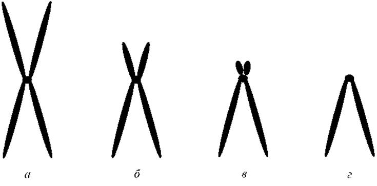
Fig. 1.10. Types of metaphase chromosomes:
a — metacentric; b — submetacentric; c — acrocentric; d — telocentric
Based on the centromere position, Human chromosomes are divided into 3 types (Figs. 1.10, 1.11):
a) metacentric — with equal arms (constriction in the middle);
b) submetacentric — one arm is longer than the other;
c) acrocentric — one arm is extremely short.
In human pathologies, telocentric (single-armed) chromosomes may occasionally be observed.
When specialized differential staining is applied, chromosomes acquire a banded appearance. Each chromosome exhibits a specific pattern of dark and light bands (an "individual portrait"), which allows for its identification under light Cell/15.html">Microscopy. This pattern is determined by the varying Nucleotide Composition of chromosomal regions, as well as The ratio of euchromatin to heterochromatin. Euchromatin is decondensed Chromatin containing a high density of actively transcribed genes. Heterochromatin is condensed chromatin containing inactive genes and non-coding highly repetitive DNA sequences.
As an example, Fig. 1.12 shows the cytogenetic map of chromosome 2. In accordance with the current international cytogenetic nomenclature (ISCN, 1995), the short arm of a chromosome is designated by the letter p, the long arm by q, and the centromere by cen. Each arm is divided into regions designated by Arabic numerals starting from the centromere towards the telomere. The boundaries between these regions are defined by specific morphological chromosomal markers. Each region is further subdivided into bands. Bands are chromosomal segments that clearly differ from adjacent ones in staining intensity (when differential chromosome staining is used). Their numbering also begins with the band closest to the centromere. Thus, the symbol 2p13 denotes the region located in the 3rd band of region 1 on the short arm of chromosome 2 (read as "2-p-one-three"). Subbands may be identified within certain bands. To designate them, an additional number is added. For example, the symbol 2p13.1 denotes the region located in the 1st subband of the 3rd band of region 1 on the short arm of chromosome 2 (read as "2-p-one-three-one").
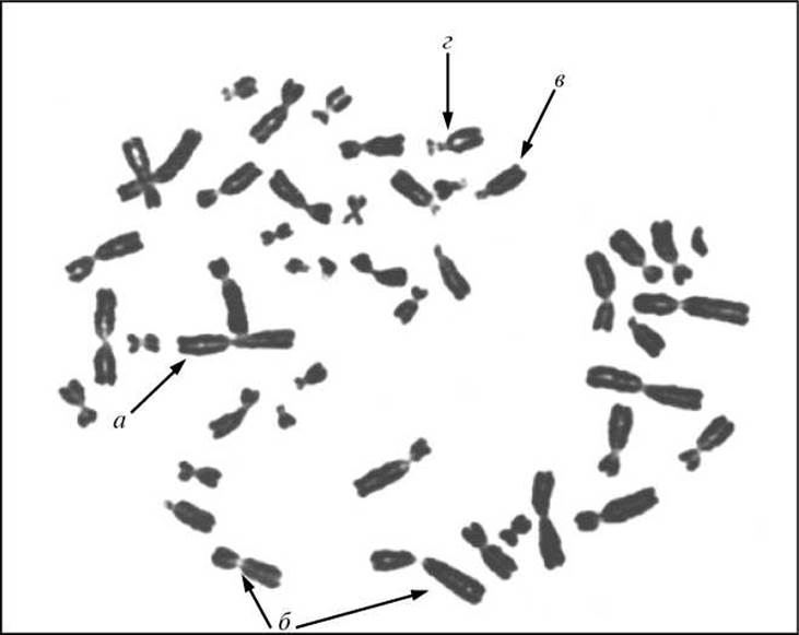
Fig. 1.11. Various types of chromosomes in a human metaphase plate:
a — metacentric; b — submetacentric; c — acrocentric; d — acrocentric, with a clearly visible satellite on the short arm
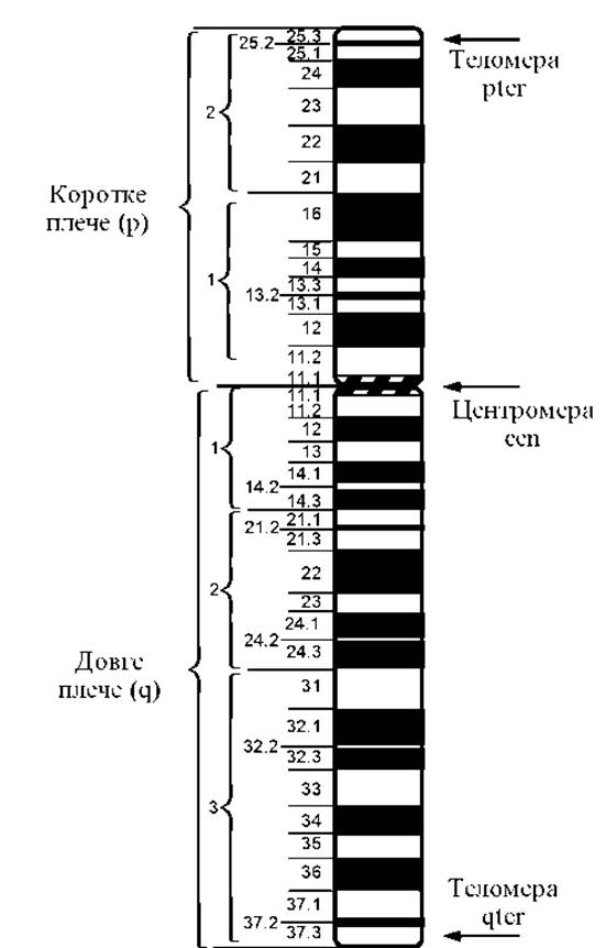
Fig. 1.12. Cytogenetic map of chromosome 2
The centromeric and most terminal telomeric regions of each chromosome arm are designated by the symbols cen and ter, respectively. For instance, the designation 17pter indicates the most distal (terminal) region of the short arm of chromosome 17.
Last update: 11/08/2026
Editorial and Educational Adaptation: This material has been compiled based on the primary/original source text. The project team performed an editorial review, corrected technical inaccuracies, structured sections, and adapted the content for an educational format.
What was processed:
- elimination of formatting defects (OCR errors, structural breaks, corrupted characters);
- editorial organization of content;
- standardization of terminology in accordance with academic sources;
- verification of factual statements against the original source text.
All mentions of the author, publication year, and origin of the primary text have been preserved in accordance with the source.