Medical Genetics - V. M. Zaporozhan 2005
Methods for Diagnosing Hereditary Diseases
Cytogenetic Methods
Molecular-Cytogenetic Methods
These Methods combine traditional cytogenetic techniques with molecular genetic technologies. The primary approach is fluorescence in situ Hybridization (FISH).
The technique involves in situ hybridization (i.e., directly on a Microscope slide preparation) of DNA probes labeled with fluorescent Dyes to metaphase or interphase Chromosomes.
Class="center">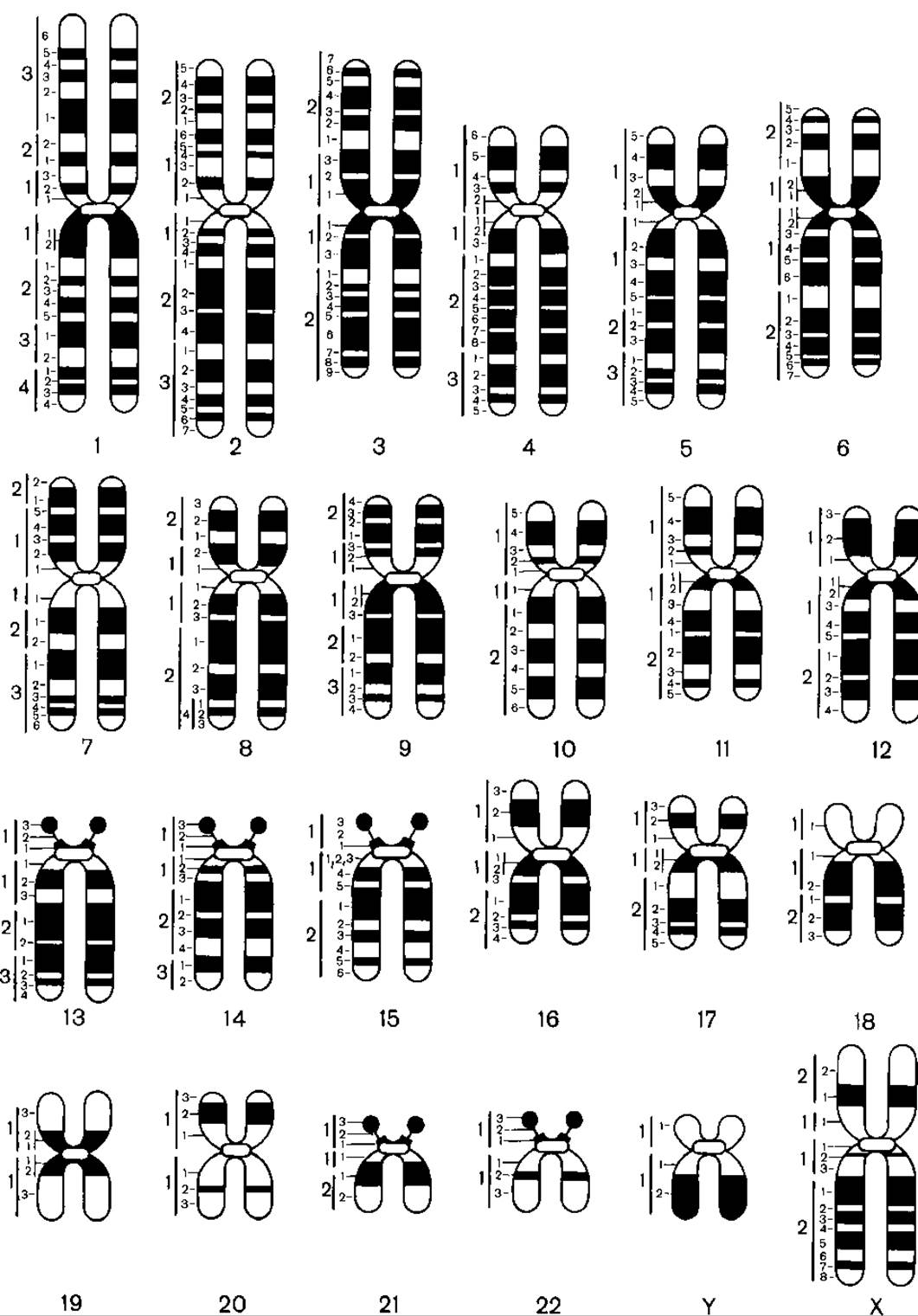
Fig. 10.2. Schematic representation of differential G-banding of chromosomes
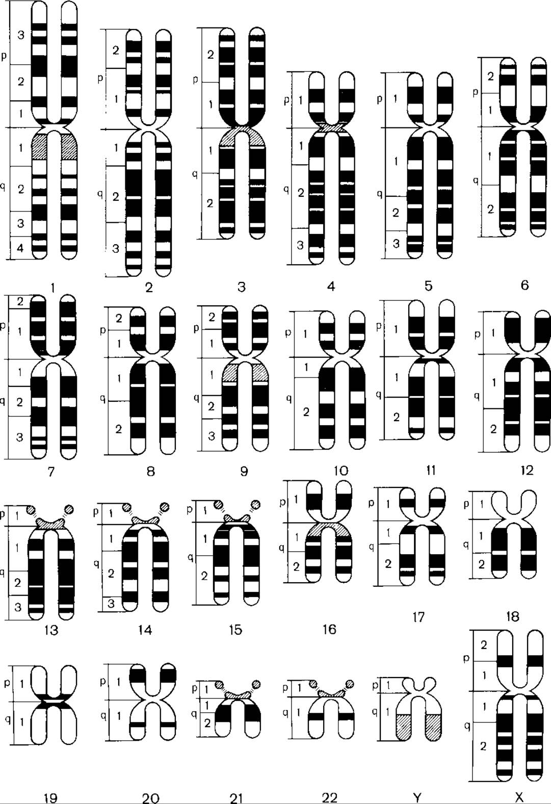
Fig. 10.3. Generalized schematic representation of differential chromosome banding using Q-, C-, and R-methods
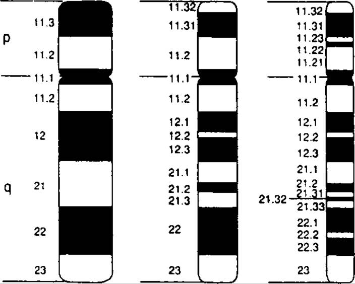
Fig. 10.4. Differential banding of chromosome 18 at various stages of Condensation (right: a metaphase chromosome with a resolution level of 400 bands per haploid set; middle and left: prometaphase chromosomes with resolution levels of 550 bands and 850 bands per haploid set, respectively)
A probe is a single-stranded DNA segment complementary to a specific Gene or chromosomal region. Probes can be genomic DNA sequences, artificially synthesized fragments, or sequences cloned using bacterial Plasmids or other methods. For the FISH method, probes are labeled with biotin or digoxigenin, which subsequently bind fluorescent dyes.
The procedure consists of the following steps:
1. Preparation of metaphase or prometaphase chromosome slides.
2. Treatment of the slide with alkali, which induces DNA Denaturation, breaking the Hydrogen Bonds between the two DNA strands.
3. Application of DNA probes in droplets onto the slide. The probe anneals to complementary chromosomal DNA sequences in a process known as hybridization. Hybridization is the joining of Nucleic Acids from different sources based on complementary base pairing (in this case, hybridization of the patient's DNA with the probe DNA).
4. Washing the slide to remove excess probe. The hybridized probes remain fixed to the chromosomes. The preparation is then treated with fluorescent dyes that attach to biotin or digoxigenin. Common dyes include rhodamine (emitting red fluorescence) or fluorescein isothiocyanate (emitting green fluorescence).
5. Examination of the slide under a fluorescence microscope. Chromosomal regions where hybridization has occurred and where the probes are localized exhibit specific fluorescence.
Today, the FISH method is also used to analyze chromosomes in non-dividing Cells during interphase (Fig. 10.5).
In clinical genetics, the method is utilized for the rapid Diagnosis of Chromosomal Disorders associated with abnormal chromosome numbers in interphase nuclei, as well as for detecting microdeletions,
microduplications, and complex chromosomal rearrangements that are rarely identified using standard cytogenetic techniques.
Various types of probes have been developed:
a) Locus-specific (gene-specific) probes. These are used to diagnose microdeletions or microduplications. For example, probes specific to a region on the long arm of chromosome 15 are used to diagnose Angelman and Prader–Willi syndromes.
Such probes are also used for chromosome mapping. In some cases, these probes can be generated from mRNA isolated from Tissues;
b) Telomere probes. A complete set of telomere probes has been synthesized for all 24 Human chromosomes (22 autosomes plus the X and Y chromosomes). The Use of these probes has enabled the detection of subtle subtelomeric deletions and translocations in a subset of children with previously unexplained intellectual disability;
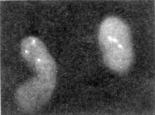
Fig. 10.5. FISH method (interphase cells from a patient with Down syndrome, treated with probes specific to chromosome 21; three fluorescent signals are visible)
c) A set of probes targeting different loci of the same chromosome. When applied simultaneously, these allow an entire chromosome to be "painted." This method enables the diagnosis of translocations involving small chromosomal segments and helps determine THE ORIGIN OF ring or marker chromosomes;
d) probes specific to the centromeres of certain chromosomes. For instance, probes targeting the centromeres of chromosomes 13, 18, 21, X, and Y have been developed. They are utilized for rapid Prenatal Diagnosis of the most common chromosomal syndromes in interphase cells from chorionic villi or Amniotic Fluid. The Principle of the method is identical to that used for metaphase spreads. A preparation of amniotic fluid cells or chorionic villi is made on a Glass slide, denatured with alkalis, and then treated with centromere-specific probes for a given chromosome (e.g., chromosome 21). Normally, somatic cells contain two copies of chromosome 21. Under a fluorescence microscope, two distinct fluorescent spots corresponding to the respective color will be visible in The Nucleus. If the fetus has Down syndrome, three spots will be observed in the nucleus (Fig. 10.5). This method is straightforward and rapid, taking only a few hours.
Modifications of the FISH Method
Reverse painting. In some cases, cells may exhibit extra chromosomal fragments such as ring or marker chromosomes. To determine their origin, the chromosome is isolated, and its DNA is amplified using the Polymerase Chain Reaction (PCR). NUCLEOTIDES labeled with the radioactive isotope 32P are used for Amplification. The resulting radiolabeled DNA is then hybridized with chromosomes from a normal metaphase spread. The Water/144.html">Origin of the additional segment is identified by matching it with the chromosome to which the labeled DNA hybridized.
Comparative genomic hybridization (CGH). This technique is widely used in Cancer genetics to detect chromosomal regions within tumors that have undergone amplification (indicating oncogene activation) or deletion (indicating the loss of a corresponding tumor suppressor gene). The principle involves isolating DNA from a tumor (the "test DNA"), amplifying it via PCR, and labeling it with a fluorescent dye. DNA isolated from normal tissue is labeled with a different dye. Next, a metaphase or prometaphase spread is prepared according to standard FISH protocols. A mixture of the test and normal DNA is then hybridized to the chromosomes. Regions of the tumor DNA where deletions (gene loss) or amplifications (gene copy number gains) have occurred are identified by shifts in fluorescence intensity.
Multiplex fluorescence in situ hybridization (multicolor spectral karyotyping / M-FISH). The FISH method employs sets of probes directed against various loci across all chromosomes, with each probe labeled with different fluorescent dyes. The karyotype is treated with this cocktail of probes, giving each chromosome pair unique spectral characteristics. Following computer-assisted spectral analysis, each chromosome pair is rendered in a distinct pseudo-color. Because every pair has a unique color, even the most complex Chromosomal aberrations can be easily identified in such a karyotype. This method is predominantly used in cancer genetics as it allows for the precise characterization of complex structural rearrangements occurring in tumor cells. Its widespread application in clinical genetics is limited primarily by its high cost.
Last update: 11/08/2026
Editorial and Educational Adaptation: This material has been compiled based on the primary/original source text. The project team performed an editorial review, corrected technical inaccuracies, structured sections, and adapted the content for an educational format.
What was processed:
- elimination of formatting defects (OCR errors, structural breaks, corrupted characters);
- editorial organization of content;
- standardization of terminology in accordance with academic sources;
- verification of factual statements against the original source text.
All mentions of the author, publication year, and origin of the primary text have been preserved in accordance with the source.